
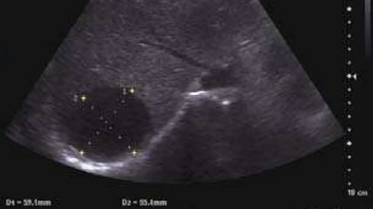
Seeing "liver cyst" or "hepatic cyst" on an ultrasound or CT report can send your mind straight to the worst place. The reassuring truth is that the large majority are simple cysts: harmless fluid-filled sacs that are not cancer, do not spread and, in most people, never need treatment. They are among the most common things found in the liver, almost always by chance. This article explains what a liver cyst is, how radiologists tell a simple one from one that needs a closer look, and what follow-up involves.
What does "liver cyst" mean?
A liver cyst is a fluid-filled sac inside the liver, like a tiny water balloon in the tissue. Most are "simple" cysts: a very thin wall, clear fluid, nothing solid inside and no connection to the bile ducts, the channels that carry bile out of the liver. They are thought to form early in life from small bile-duct structures that never joined the drainage system, then slowly fill with fluid, which is why they are found more often with age. A few percent of adults have one on imaging, often just one.
A simple cyst looks the same way each time:
- On ultrasound, a round, completely black (anechoic) area with a wall too thin to see and brighter tissue behind it
- On CT, a smooth, water-dark area that does not light up after contrast dye (a liquid injected into a vein)
- On MRI, plain-water behavior: very bright on T2-weighted images, with no enhancement
When all of these boxes are ticked, radiologists are confident the cyst is benign and usually no further tests are needed, which is also how the American College of Radiology's guidance on incidental liver findings treats it.
What causes liver cysts?
For most people there is no cause to find: simple cysts are a developmental quirk, not the result of anything you ate, drank or did. They become more common with age and are more frequent, and tend to be larger, in women, so hormones are suspected to play a part. They are unrelated to kidney cysts, though both are common and can appear on the same scan.
When a liver cyst is not a simple cyst
Some conditions look like a cyst or contain cysts. Radiologists look for thick walls, internal partitions, solid nodules, calcium and debris:
- Polycystic liver disease: many cysts, often inherited and sometimes alongside polycystic kidney disease
- Hydatid (echinococcal) cyst: a tapeworm cyst, picked up mostly where people live closely with sheep and dogs, including parts of the Mediterranean and Middle East. It can show a double wall, smaller "daughter" cysts or a calcified rim
- A cyst that has bled or become infected, or an abscess: cloudy fluid or debris, often with pain or fever
- Cystic tumor: rare. A biliary cystadenoma has thick partitions or nodules and is usually removed because it can, rarely, turn cancerous
Is it serious?
Usually not. A simple liver cyst has no potential to become cancer, the same reassurance our guide on whether a cyst can be cancerous gives for cysts in general. Doctors look harder at a cyst with a thick wall, partitions, a solid nodule or calcium, at one that grows on a repeat scan, and at one that comes with symptoms.
Contact your doctor promptly if a known cyst comes with sudden severe pain in the upper right abdomen, fever with abdominal pain, or yellowing of the skin or eyes. Interpretation always depends on symptoms, tests and history, not on the scan alone.
Symptoms
Usually none. Most liver cysts are silent, found by chance, and liver blood tests are usually normal. A cyst causes symptoms mainly when it is large, commonly several centimeters across: a dull ache or fullness in the upper right abdomen, feeling full after small meals, bloating or nausea. Rarely there is a complication, such as bleeding into the cyst, infection or pressure on a bile duct.
How is it diagnosed and followed up?
Ultrasound is often the first test. A CT or MRI may confirm it, and MRI is the most reliable for small or doubtful spots. What follows depends on the cyst and on you:
- Clearly simple cyst: usually nothing further
- "Too small to characterize": a dot under about 1.5 cm may be too small for CT to label with certainty. With no known cancer or liver disease it is very often a tiny cyst and is left alone, while a history of cancer may prompt an MRI
- Atypical or complex cyst: an MRI with contrast, perhaps a repeat scan after a few months, blood tests and a liver specialist
- Many cysts: a look at the kidneys, the family history and liver and kidney function
Follow-up recommendations vary by patient, and your own doctor knows your history.
Treatment options
For most people, none: a simple cyst without symptoms is left alone, and no diet, supplement or exercise is proven to shrink one. Treatment is considered only for a large cyst that causes real symptoms or looks worrying:
- Needle drainage with sclerotherapy: the fluid is drained under imaging guidance and a substance such as alcohol is injected to seal the lining, because draining alone often lets the cyst refill
- Laparoscopic fenestration: keyhole surgery removes part of the wall so fluid stops collecting, usually with fewer recurrences than draining alone
- Other causes: removal for a suspected cystic tumor, anti-parasite medicine with a procedure for a hydatid cyst, and for polycystic liver disease options that follow the symptoms
Why a second read can help
The line between a simple cyst and something that needs a closer look rests on subtle details. Is a faint line inside the cyst a partition or an artifact? Is a tiny dark dot a cyst or a small hemangioma, the harmless blood-vessel growth in our article on hepatic hemangioma? An expert second read can confirm which side of that line your cyst sits on and put it in plain words. If you would like an online radiology second opinion, DocOrbit lets you send the study from home, and a subspecialist radiologist writes a structured report you can share with your own doctor. Our guide to when to get a second radiological opinion can help you decide, and the abdomen and pelvis MRI second opinion page shows what a full review covers.
Can a liver cyst turn into cancer?
A simple liver cyst does not turn into cancer: it is a benign fluid-filled sac with a thin wall and no solid tissue. The concern is with rare cysts that look complex, with thick walls, partitions or solid areas, which is why radiologists note those features and sometimes suggest an MRI or a follow-up scan.
How big does a liver cyst have to be to cause pain?
There is no single size, and many large simple cysts cause no symptoms at all. Pain, when it comes, usually starts once a cyst is several centimeters across and presses on nearby structures, as a dull ache or fullness in the upper right abdomen.
Do liver cysts need to be drained or removed?
Almost never. A simple cyst without symptoms is left alone. Draining or surgery is considered only when a large cyst causes pain or pressure, becomes infected or bleeds, or looks like something other than a simple cyst.
Key takeaways
- Most liver cysts are simple cysts: benign, fluid-filled, not cancer and common, especially with age
- A clearly simple cyst usually needs no treatment and no follow-up
- Thick walls, partitions, solid nodules, calcium, growth or symptoms prompt a closer look, often with an MRI
- Sudden severe pain in the upper right abdomen, fever with pain or yellow skin needs prompt attention
If the rest of your report reads like another language, our free radiology report explainer rewrites it in everyday words. And to go through the images with a specialist, you can ask a radiologist online through DocOrbit and take the answers back to your own doctor.
Can I ask a radiologist about my liver scan report online?
Yes. You upload your images from home, with no CD to post and no second trip, and a subspecialist radiologist re-reads the study and writes a structured report you can hand to your own doctor within 24 to 48 hours. You can message the radiologist with questions afterwards, including whether the cyst on your report is a simple one or needs a closer look.
This article is for general information only and is not medical advice. Always discuss your imaging results and any next steps with a qualified physician.