Few lines on a radiology report cause more quiet worry than the word "mass" or "lesion" sitting next to "liver." If yours mentions a hepatic hemangioma, here is the reassuring part first: it is the most common non-cancerous growth found in the liver, it is simply a tangle of blood vessels, and in the overwhelming majority of people it never causes a problem or needs treatment. This article explains what a hemangioma actually is, why it forms, when it occasionally matters, and what follow-up usually looks like.
What does "hepatic hemangioma" mean?
A hepatic hemangioma is a benign collection of widened, blood-filled vessels in the liver. Think of it less like a tumor in the frightening sense and more like a small, slow pool of blood vessels that formed differently as the liver developed. The word "hemangioma" simply combines the terms for blood vessel and benign growth. It describes the structure, not a disease process.
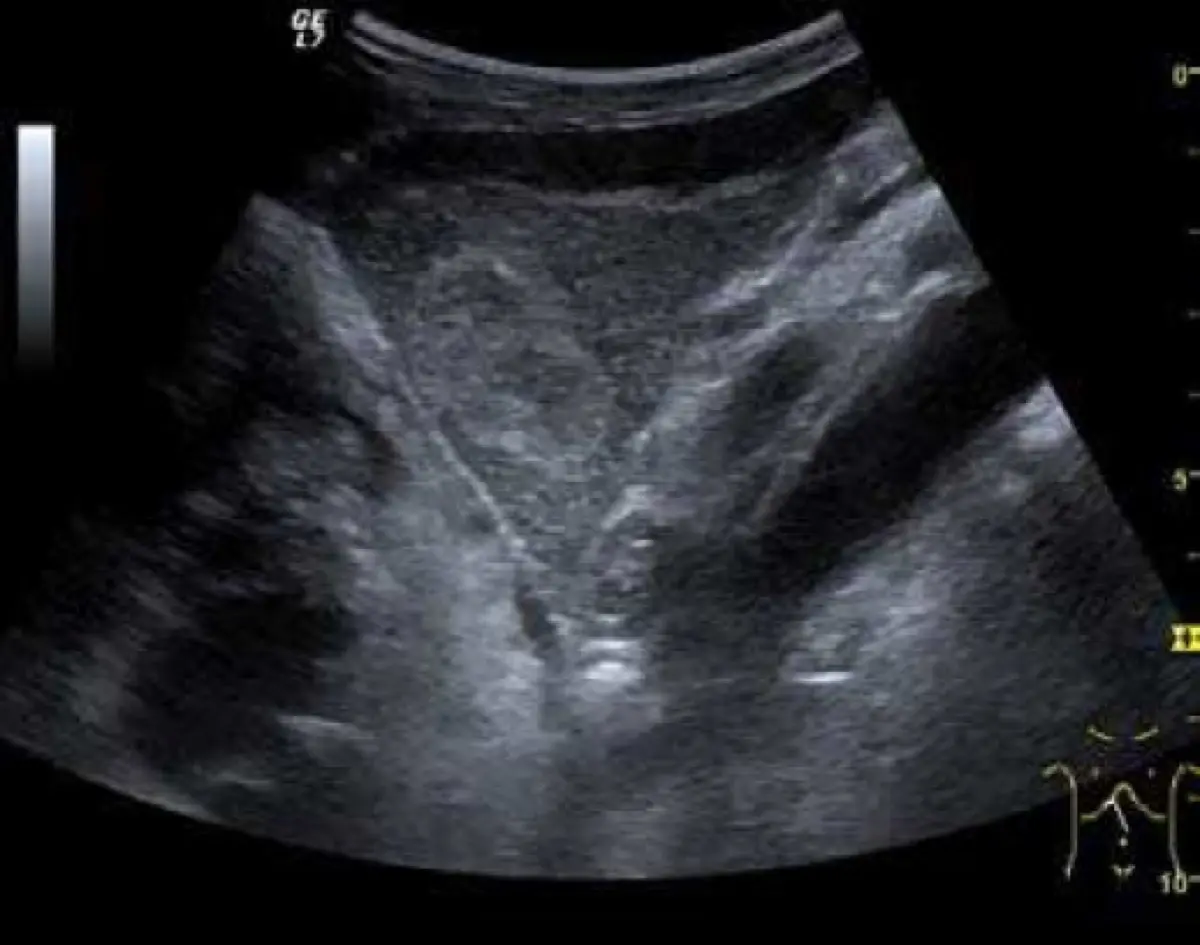
It is usually discovered by accident, when a scan is done for something else entirely: abdominal pain, gallstones, a routine check, or a follow-up for an unrelated issue. On imaging, radiologists look for a recognizable pattern:
- On ultrasound, a classic small hemangioma is often a well-defined, bright (echogenic) spot with smooth edges
- On MRI, it tends to be strikingly bright on T2-weighted images, an appearance radiologists sometimes describe as a "light bulb" sign
- On CT or MRI with contrast, it characteristically fills in from the outer edge toward the center over successive image phases, a behavior radiologists find reassuring
When a lesion shows these textbook features, a confident diagnosis can often be made on imaging alone, without any need for a biopsy.
Common causes
Hemangiomas are best understood as something you were essentially born with, not something you developed from a habit or exposure. The usual themes:
- Congenital origin: most are thought to be a small vascular malformation present from birth that simply grows very slowly over the years
- Hormonal influence: they are more common in women, and estrogen (during pregnancy or with some hormone medications) can make them enlarge somewhat
- Age of discovery: they are most often picked up between the ages of 30 and 50, largely because that is when people start having abdominal scans for other reasons
Importantly, hemangiomas are not caused by alcohol, diet, hepatitis, or fatty liver. If you are also being told about fat in the liver, that is a separate finding. Our guide to fatty liver disease and the role of MRI covers that one on its own.
Is it serious?
For the vast majority of people, a hemangioma is an incidental finding: noted, characterized, and then safely ignored. The reassurance is genuine: a hemangioma has no known potential to become cancer, and most never grow in a meaningful way.
There are a few situations where a hemangioma earns a closer look:
- Giant hemangiomas: those larger than about 5 cm, and especially over 10 cm, are more likely to cause symptoms or warrant monitoring
- An atypical appearance: if the imaging features are not classic, the priority shifts to confirming it really is a hemangioma and not something that mimics one
- A background of cancer or chronic liver disease: in someone with a known cancer or cirrhosis, radiologists are appropriately more careful before calling any liver spot benign
This is the same principle behind investigating any incidental spot carefully rather than assuming the worst. We describe the same calm, stepwise logic for a lung nodule found on a CT scan.
Symptoms
Most hemangiomas cause no symptoms at all, which is exactly why they are usually found by surprise. When a hemangioma is large, it can occasionally produce vague upper-right abdominal discomfort, a sense of fullness, or feeling full quickly when eating. This happens simply because its size presses on neighbouring structures. Pain that is sudden or severe is unusual and deserves prompt medical attention rather than watchful waiting.
How is it diagnosed and followed up?
The goal of the workup is confidence, not intervention. When the appearance is classic on ultrasound, CT, or MRI, no further testing may be needed. When there is any doubt, a contrast-enhanced MRI is often the most useful next step, because the way a hemangioma fills with contrast over time is highly characteristic.
Follow-up imaging is reserved for lesions that are large, atypical, or found in a higher-risk liver, and it usually means a repeat scan some months later. Biopsy is generally avoided for a suspected hemangioma, because sampling a mass made of blood vessels carries an unnecessary bleeding risk and is rarely needed when imaging is convincing.
Treatment options
For a typical hemangioma, the treatment is observation. In practice, that often means doing nothing at all. The small minority that cause genuine problems (a giant lesion with persistent symptoms, or the rare case of bleeding) may be managed by a specialist with options such as surgical removal or blocking the lesion's blood supply (embolization). These are exceptions, not the rule, and the decision always weighs the lesion against the risks of any procedure.
Why a second read can help
Most of the anxiety around a liver lesion comes down to a single question: is the radiologist sure it is a hemangioma and not something else? That confidence rests entirely on reading the imaging features correctly. Benign liver lesions can occasionally mimic, or be mimicked by, more important ones. An independent expert second read of your MRI or CT can confirm the diagnosis, or flag where another opinion is warranted, and give you something concrete to share with your own doctor. Services like DocOrbit make that kind of second radiological opinion straightforward, which is reassuring precisely because confirming "benign" is as valuable as catching something missed.
Is a hepatic hemangioma dangerous?
For the large majority of people, no. A hepatic hemangioma is a benign tangle of blood vessels, not a cancer, and most cause no symptoms and need no treatment. The uncommon exceptions are very large hemangiomas that press on nearby organs or, very rarely, bleed. The appearance on imaging, your symptoms, and your medical history together decide whether it needs any attention at all.
Can a liver hemangioma turn into cancer?
No. A hemangioma is a malformation of blood vessels and has no known potential to become liver cancer. The main reason radiologists pay attention to one is to be confident it really is a hemangioma and not a different lesion that happens to look similar, which is why imaging features matter so much.
Do hepatic hemangiomas need to be removed?
Almost never. The standard approach for a typical hemangioma is simply to leave it alone. Removal or other procedures are reserved for the small number that grow very large, cause persistent pain or pressure symptoms, or bleed, which is rare. Most people live their whole lives with a hemangioma and never need anything done about it.
What causes a hemangioma on the liver?
Hepatic hemangiomas are thought to be present from birth as a small vascular malformation that the liver simply forms differently. They are not caused by diet, alcohol, or infection. They are more common in women and can enlarge under the influence of estrogen, such as during pregnancy or with some hormone medications, though most stay stable for years.
Should a liver hemangioma be rechecked?
When the imaging features are classic, many hemangiomas need no follow-up at all. A repeat scan in several months is more likely when the lesion is large, looks atypical, or is found in someone with a history of cancer or chronic liver disease, where confirming the diagnosis matters more. Your doctor tailors this to your situation rather than a fixed rule.
Key takeaways
- A hepatic hemangioma is the most common benign liver growth: a harmless tangle of blood vessels, not a cancer
- Most are found by accident, cause no symptoms, and need no treatment
- Classic imaging features often allow a confident diagnosis without a biopsy
- Large, atypical, or higher-risk cases are the ones that get follow-up or a closer look
- Confirming a benign diagnosis is exactly where a careful or second read adds peace of mind
This article is for general information only and is not medical advice. Always discuss your imaging results and any next steps with a qualified physician.