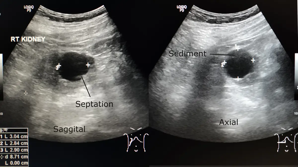
Almost never, but yes — a small number of cysts do turn out to be cancerous, and radiologists work through a specific set of features to tell a harmless cyst from one that needs a closer look.
The overwhelming majority of cysts, simple fluid-filled sacs found in the kidneys, ovaries, breasts, and liver, are entirely benign and need no treatment. What decides whether a cyst is one of the rare exceptions is not which organ it is in, but what it looks like on the scan: a thin or thick wall, truly clear fluid or not, and whether anything solid is growing inside it. That is what this article walks through.
What separates a "simple" cyst from one that gets a second look
Picture a simple cyst as a water balloon: a thin, smooth wall around clear fluid, with nothing solid inside. On ultrasound or MRI, that shows up as a round, uniform pocket with no internal structure, by far the most common pattern, and about as reassuring as a finding gets.
A cyst that earns a closer look resembles a balloon with a small pebble inside it, or one divided into compartments by thin internal walls. Radiologists have specific words for these features:
- Septations — thin walls dividing the fluid into compartments. A few thin ones are usually fine; thick or numerous ones raise a flag.
- Mural nodule — a small solid lump attached to the cyst wall, not free-floating debris.
- Wall thickening — a wall that is no longer thin and smooth, but irregular or thick instead.
- Internal blood flow — on Doppler ultrasound, a mode that shows moving blood as color, a solid part with its own blood supply is taken more seriously.
None of these features alone means cancer; they simply shift a cyst from watch-and-forget to worth another look.
Which organs get flagged more often
The threshold for concern differs by organ:
- Kidney — kidney cysts are graded on the Bosniak scale, from I, simple and essentially never cancerous, to IV, mostly solid and usually removed. Most kidney cysts found on ultrasound are Bosniak I and simply left alone.
- Ovary — most ovarian cysts before menopause come and go with the menstrual cycle. A complex-looking cyst after menopause is taken more seriously than the same appearance at 25.
- Breast — a simple breast cyst on ultrasound is benign by definition. A complicated or complex one, with internal echoes or a solid part, may prompt a biopsy to be sure.
- Pancreas — most pancreatic cysts found incidentally on CT or MRI are IPMNs, usually watched rather than removed unless they show specific worrisome features.
- Liver — simple liver cysts are extremely common and essentially never cancerous.
What happens if your cyst looks complex on the scan
A complex cyst is not treated as cancer by default. The usual next step is repeat imaging after a set interval, often months rather than weeks, to see whether it has changed, sometimes alongside a referral to a specialist, a follow-up MRI, or a needle sample of any solid portion.
Before choosing between watching a complex cyst and going straight to biopsy, some patients get an online radiology second opinion on the actual images, not just the typed report, especially when the first read already flagged something ambiguous. A service like DocOrbit can connect you with a subspecialist radiologist to look at the scan directly.
Does a painful cyst mean it is more likely to be cancerous?
No. Pain from a cyst usually relates to its size, location, or a rupture or torsion, the cyst twisting on itself, mainly a risk with larger ovarian cysts, not to malignancy. Sudden, severe pain can be a genuine emergency and needs urgent evaluation, but pain alone does not shift the odds toward cancer.
Can a cyst turn into cancer over time?
A true simple cyst does not transform into cancer. Rarely, a cyst that already had complex features on the first scan can develop a cancer within it over months to years, which is why complex cysts are monitored instead of ignored. This is best documented in certain ovarian cysts and pancreatic IPMNs, not in simple cysts anywhere in the body.
Is a growing cyst automatically more concerning?
Growth is one reason a cyst gets watched more closely, but it is not proof of anything by itself. Many benign cysts grow slowly over years without ever becoming a problem. What matters more is whether new complex features, like a new solid nodule or new blood flow, appear alongside that growth.
How do I have my ultrasound report reviewed by another radiologist online?
You upload your images or ultrasound report from home, no CD and no extra trip to a clinic, and a subspecialist radiologist reviews it and writes a structured report you can share with your own doctor, usually within 24 to 48 hours. You can message the radiologist afterward with questions about what the report means. If you want an answer sooner, Orbius, DocOrbit's AI radiology agent, gives a preliminary structured read in minutes, with the option to add a signed specialist report later.
Key takeaways
- The large majority of cysts, in any organ, are simple, benign, and need no treatment.
- What matters is appearance, not location: a thin wall and clear fluid reassure; septations, a solid nodule, or a thickened wall get a closer look.
- Different organs use different systems, Bosniak for the kidney, O-RADS for the ovary, BI-RADS for the breast, to decide on follow-up.
- A cyst that changes on repeat imaging is watched more closely, not automatically treated as cancer.
This article is for general information only and is not medical advice. Always discuss your imaging results and any next steps with a qualified physician.
