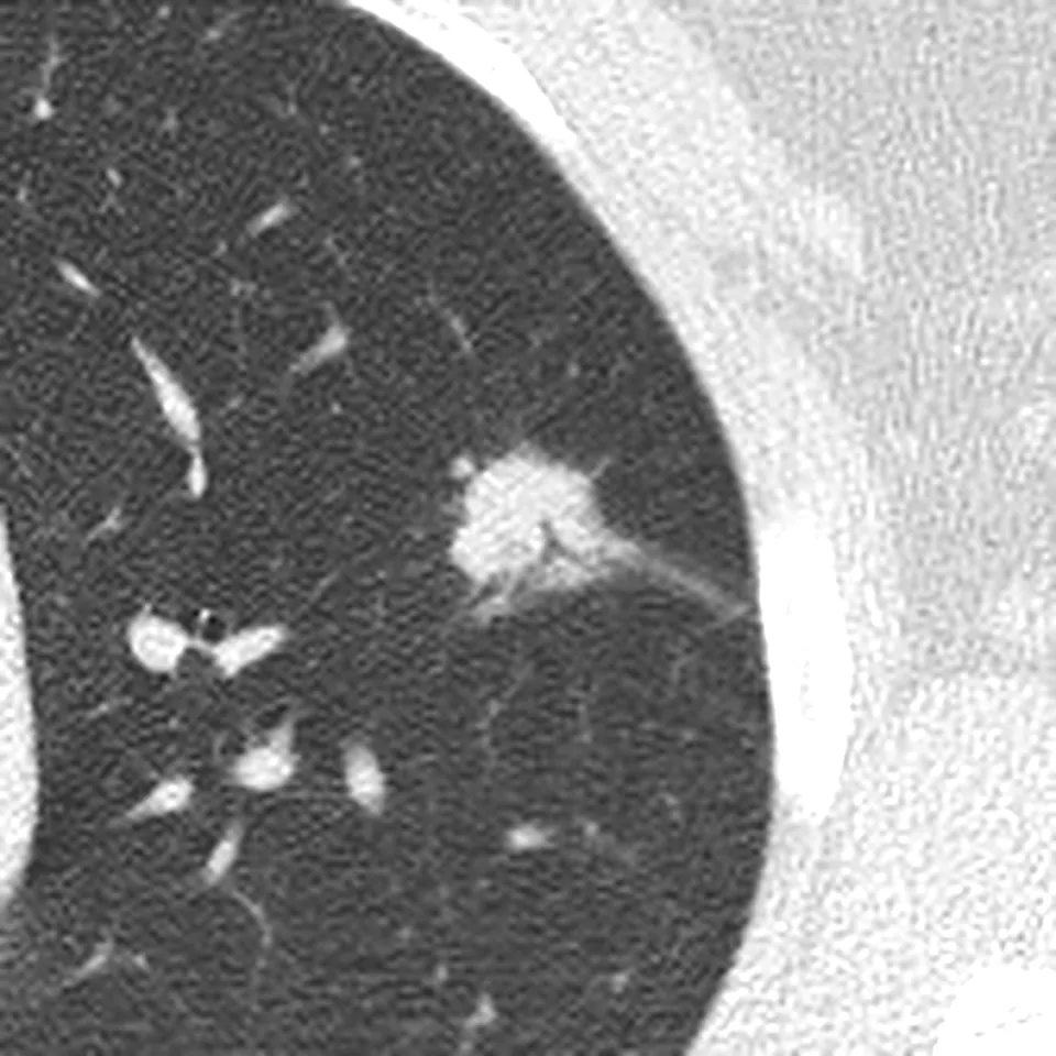
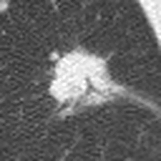
One chest CT, read both ways: by Orbius, our AI, and by a board-certified radiologist. These are two separate options, and you choose the one you need. The scan is real and public; the patient and both reports are fictional.
Option 1 · in minutes
The Orbius AI report
Orbius reads the full study and writes a structured report, from $20. No doctor reviews it, so it is not a diagnosis.
Orbius AI report · not signedCT chest · low-dose, no contrast
Indication
Screening in a 68-year-old former smoker
Findings
An 18 mm solid nodule in the upper left lung, with spiculated edges.
Spiculated means spiky rather than smooth. Together with the size, the spiky edge is the reason to check this nodule promptly.1
No enlarged lymph nodes in the chest.
Mild emphysema at the top of both lungs, consistent with the smoking history.
No earlier chest scan on file to compare with, so there is no way to tell whether the nodule is new.
Conclusion
A single nodule with irregular, spiky edges. Its size and shape, with nothing earlier to compare against, are the reasons to check it promptly rather than wait and see.
Next step2
PET-CT in the next few weeks, and a radiologist's review of this study
Option 2 · in 24 to 48 hours
The radiologist's second opinion
A board-certified chest radiologist reads the images, writes the report and signs it. This is the full report you receive, from $60.
Lung cancer screening, first scan. Former smoker, 40 pack-years, stopped 6 years ago. Second opinion requested on the original report.
Technique
Low-dose CT of the chest without contrast, from the top of the lungs to the adrenal glands. 1.25 mm images in lung and soft-tissue windows, with coronal and sagittal views.
Comparison
No earlier chest CT available. The original report of this scan (21 Apr 2026) was reviewed.
Findings
Lungs. Solid nodule in the left upper lobe, 18 × 15 mm (series 2, image 190), with spiky edges (spiculated) and a thin strand pulling on the nearby lining of the lung (pleural tag).
Needs prompt work-up · PET-CT
Lungs. Mild emphysema in both upper lobes. No other nodules.
Airways. The windpipe and main airways are clear.
Lymph nodes. No enlarged lymph nodes in the chest or at the roots of the lungs.
Pleura. No fluid around the lungs (no pleural effusion).
Heart and vessels. Normal heart size. Mild calcium in the heart arteries (coronary calcification).
Unrelated finding
Upper abdomen and bones. Nothing abnormal in the upper abdomen in view. Mild wear of the thoracic spine.
A single 18 mm nodule in the left upper lobe with spiky edges. Its size and shape need prompt work-up (Lung-RADS 4X).
No sign of spread to the lymph nodes or the lining of the lung.
Mild emphysema and mild coronary calcification, both common in former smokers.
Compared with your original report5
Agrees with your original report, which describes the same 18 mm nodule in the left upper lobe and advises further evaluation. This review adds three things: a risk category (Lung-RADS 4X, because of the spiky edge and the strand to the pleura), a clear next step with a time frame (PET-CT within two weeks), and the nodule outlined and measured on your images.
Recommendations
PET-CT within two weeks to see how active the nodule is.
Review by the lung team (thoracic multidisciplinary meeting) once the PET-CT is back.
If the PET-CT is unclear, a CT-guided biopsy can be considered.
Discuss the coronary calcium with your GP as part of a heart-health check.
In plain words6
Your scan shows one spot in the upper part of your left lung. Its spiky edge is the reason to look at it soon. A PET-CT will show how active it is, and there is no sign of spread. Take this report to your doctor or lung specialist to plan the next step.
Two ways to get your scan read
Choose the read you need
Two separate options. You see the exact price before you pay.
The Orbius AI report compared with a radiologist second opinion
Orbius AI reportfrom $20
Radiologist second opinionfrom $60
Who reads your scan
Orbius, our AI
A board-certified radiologist matched to your scan
What you get
A structured report, with every term explained
A signed report, compared with your original report