
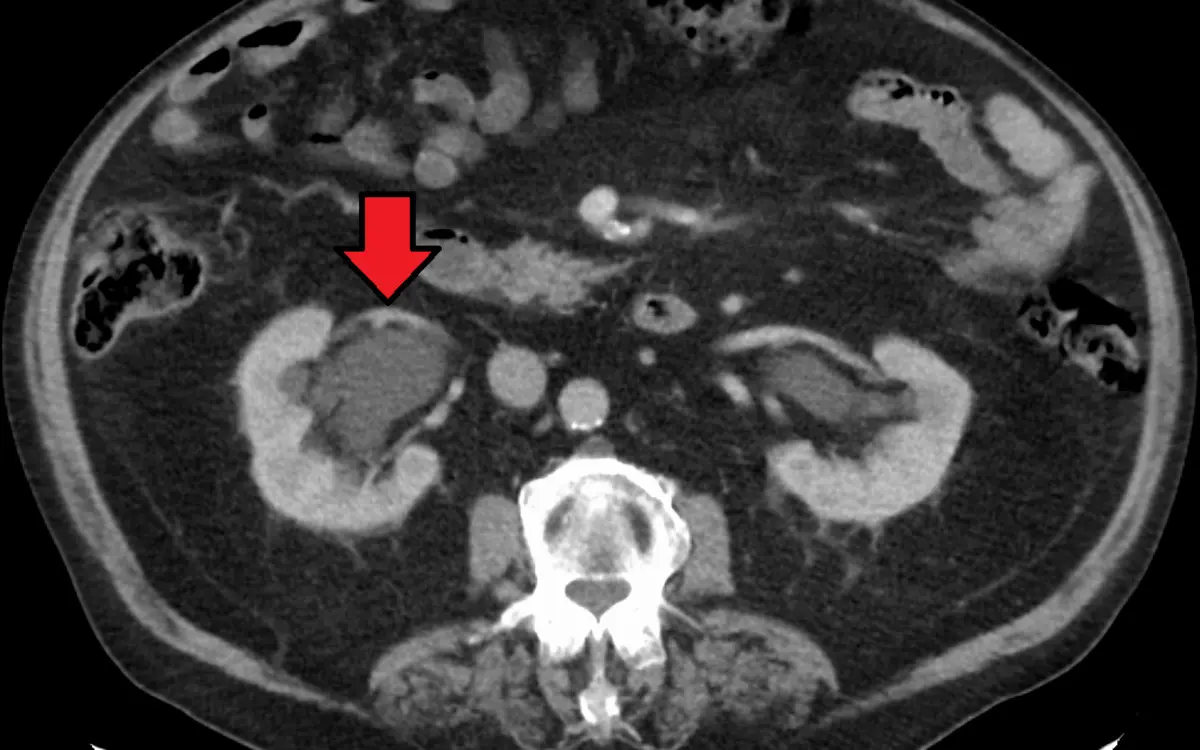
Seeing "hydronephrosis" on an ultrasound or CT report can be unsettling, mostly because nobody explains the word. The reassuring truth is that it is not a disease in itself: it simply means a kidney is holding more urine than usual and looks swollen. In many people it is mild and either settles on its own or has a simple explanation such as a full bladder or pregnancy. What matters is why the urine is backing up. Here is what the finding means, what causes it, when it needs prompt attention and how it is followed up.
What does "hydronephrosis" mean?
Hydronephrosis (from the Greek for "water" and "kidney") means swelling of the kidney's drainage system. Each kidney makes urine, gathers it in small cups called calyces, funnels it into a central basin called the renal pelvis and sends it down a thin tube, the ureter, to the bladder. When urine cannot drain freely, or flows back up, the cups and the pelvis stretch like a balloon filling with water.
Reports use several wordings for the same idea: "pelvicalyceal dilatation", "caliectasis", "pyelectasis" (a mildly widened renal pelvis) or "fullness of the collecting system". If the ureter is widened too, you may read "hydroureter" or "hydroureteronephrosis", which helps the radiologist work out where along the path the problem lies.
This is how it looks on each test:
- On ultrasound, branching black (anechoic) channels of fluid in the middle of the kidney, where there is normally a compact, bright core. It is usually the first test and uses no radiation
- On CT, a water-dark area in the center of the kidney that does not take up contrast dye (a liquid injected into a vein). CT is also better at showing the cause, such as a stone, along the ureter
- Radiologists often grade it as mild, moderate or severe. In babies and children, the Society for Fetal Urology scale from 0 to 4 is common
What causes hydronephrosis?
Causes fall into two groups: something blocks the urine on its way out, or the system is wide without a true blockage. The common ones are:
- A stone in the ureter: one of the most frequent reasons for sudden hydronephrosis, covered in our guide to kidney stones on CT and ultrasound
- A narrowing of the ureter: either where it leaves the kidney (ureteropelvic junction obstruction), often present from birth, or a scar-like stricture after an infection, an operation or earlier stones
- Trouble emptying the bladder: in men, an enlarged prostate can make urine back up into both kidneys
- Pressure from outside or a growth inside: a mass in the abdomen or pelvis, enlarged lymph nodes, a tumor of the ureter or bladder or, rarely, scar tissue around the ureters
- Reflux: urine flowing back from the bladder toward the kidney, mostly in children
- No blockage at all: a very full bladder at the time of the scan, a lot of urine being made after drinking plenty, or a kidney that simply has a roomy pelvis
- A look-alike: simple cysts next to the pelvis (peripelvic cysts, as in our kidney cyst guide) can imitate a swollen pelvis on ultrasound
Hydronephrosis in pregnancy and in babies
Mild hydronephrosis is common in pregnancy, more often on the right side. The growing uterus presses on the ureters and pregnancy hormones relax their walls. It usually causes no trouble and settles after delivery, though pain with fever deserves a prompt check.
In babies it is often spotted on the routine pregnancy ultrasound. Most mild cases settle or stay stable, but some come from a narrowing or reflux, so the ultrasound is repeated after birth and a pediatric urologist or kidney specialist guides any further tests.
Is hydronephrosis serious?
It depends on the cause, how swollen the kidney is, how quickly it developed and whether one kidney or both are affected. Doctors take it more seriously, and act faster, in these situations:
- Fever or chills with flank pain: a blocked kidney that becomes infected can turn dangerous and needs urgent drainage
- Severe pain or vomiting that will not settle
- Both kidneys affected, or only one working kidney, or passing very little urine
- Kidney blood tests that are getting worse
These are reasons to seek medical care promptly rather than wait for a routine appointment. A blockage that has been there a long time matters too, even without symptoms, which is why a swollen kidney is followed up and not simply ignored.
Symptoms
Often none. Mild or slow-growing hydronephrosis is typically silent and discovered on a scan done for another reason. When symptoms do appear they come from the cause:
- Sudden, cramping pain in the side or back that comes in waves and spreads toward the groin, usually from a stone
- A dull ache in the flank, sometimes brought on by drinking a lot
- Blood in the urine, or repeated urinary infections with fever
- A weak or slow stream, or the feeling that the bladder does not empty fully
How is it found and followed up?
Ultrasound is the usual first test. It shows the swelling and how much kidney tissue is left around it, and it is safe in pregnancy and in children. A CT of the abdomen and pelvis, often without contrast when a stone is suspected, is better at finding the cause: a stone, a mass or a narrowing along the ureter. Blood tests for kidney function (creatinine) and a urine test are almost always part of the picture.
When the scan cannot say whether the swelling is really blocking the flow, a nuclear medicine test can. A diuretic renogram (MAG3 scan) measures how quickly each kidney drains, as described in our guide to renal scintigraphy. In mild cases with no clear cause, the usual next step is a repeat ultrasound after a few weeks or months to see whether the swelling is settling, stable or growing. Follow-up recommendations vary by patient, and your own doctor knows your history.
Treatment options
Hydronephrosis itself is not treated directly. Doctors treat the cause, with the aim of letting urine drain and protecting the kidney:
- Watchful waiting: for mild cases with no symptoms, no infection and normal kidney tests, including most pregnancy-related ones
- Stones: pain relief and time for a small stone to pass, or lithotripsy or ureteroscopy for a larger one
- Infection: antibiotics and, when a blocked kidney is infected, urgent drainage with a ureteral stent (a thin tube placed from the bladder up to the kidney) or a nephrostomy tube (placed through the skin of the back directly into the kidney)
- Prostate or bladder outlet: medicines, a temporary catheter or surgery
- A narrowing: repair, for example a pyeloplasty for a ureteropelvic junction obstruction, or a stent or dilation for a stricture
- A mass or tumor: treatment aimed at that cause
No lifestyle change shrinks hydronephrosis on its own, though when stones are behind it, drinking enough fluid is the most useful habit.
Why a second read can help
A line such as "mild hydronephrosis" rests on judgment calls. Is the pelvis truly dilated, or just full because the bladder was? Is a small bright dot along the ureter a stone or a calcified vein in the pelvis (a phlebolith), which can look alike? Is it a swollen pelvis or cysts imitating one? An expert second read can settle these points and put the finding in plain words. If you would like an online radiology second opinion, DocOrbit lets you upload the study from home, and a subspecialist radiologist writes a structured report you can share with your own doctor. Our guide to when to get a second radiological opinion can help you decide, and the abdomen and pelvic CT second opinion page shows what a full review covers.
Is mild hydronephrosis serious?
Usually not. Mild hydronephrosis with no pain, no fever and normal kidney tests is common, often temporary, and is typically watched with a repeat ultrasound rather than treated. It matters more if it grows, if it comes with infection or pain, or if a cause such as a stone or a blockage is found.
Can hydronephrosis go away on its own?
Often, yes. Hydronephrosis caused by a stone that passes, by pregnancy, by a very full bladder or by a short-lived infection usually settles once the cause is gone, and many mild cases in babies improve with time. Hydronephrosis caused by a lasting narrowing or blockage does not clear until that cause is treated.
Can hydronephrosis damage the kidney?
It can if urine stays backed up for a long time, because the pressure slowly thins the kidney tissue and lowers its function. That is why doctors look for the cause, relieve a true blockage and use repeat scans and kidney blood tests to keep watch. A kidney that is unblocked in good time often recovers well.
Key takeaways
- Hydronephrosis means a kidney is swollen with urine. It is a finding rather than a diagnosis, and the cause is what counts
- Mild cases are common, often temporary and usually watched with a repeat ultrasound
- Stones, a narrowing, prostate trouble and pregnancy are among the usual causes
- Fever, severe pain, both kidneys affected or very little urine need prompt medical care
Your report probably holds more than this one finding. Our free radiology report explainer rewrites the whole thing in everyday words. And to go through the images with a specialist, you can ask a radiologist online through DocOrbit and take the answers back to your own doctor.
Can I ask a radiologist about my kidney scan report online?
Yes. You upload your images from home, with no CD to post and no second trip, and a subspecialist radiologist re-reads the study and writes a structured report you can hand to your own doctor within 24 to 48 hours. You can message the radiologist with questions afterwards, including whether the swelling on your report is mild and likely to settle or needs a closer look.
This article is for general information only and is not medical advice. Always discuss your imaging results and any next steps with a qualified physician.