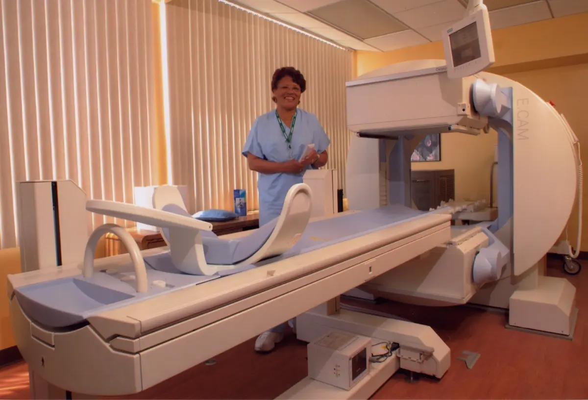
You were sent for a kidney scan, someone injected a small amount of a radioactive tracer, you lay under a large camera for half an hour, and now a report has come back talking about split function, delayed drainage, and possibly cortical scarring. Nobody explained any of it. Here is the reassuring starting point: this test exists precisely because it can answer a question no ultrasound or CT can answer, and most results are far less dramatic than the vocabulary suggests.
What a renal scintigraphy actually measures
An ultrasound or a CT shows you the shape of the kidneys: how big they are, whether they are swollen, whether a stone or a cyst is sitting somewhere. Those are pictures of structure. What they cannot tell you is how hard each kidney is working, or whether urine is genuinely getting out.
Renal scintigraphy answers those two questions. A tiny amount of a tracer is injected into a vein, the kidneys handle it the way they handle everything else in the blood, and a gamma camera records where it goes and how quickly it leaves. The output is not a photograph. It is a curve over time, plus a percentage, and that is why the report reads so unlike an ultrasound report.
There are two main flavours, and knowing which one you had explains most of the wording:
- MAG3 (or DTPA) renogram: a dynamic study. The camera films the tracer arriving, concentrating, and draining away over roughly 20 to 30 minutes. This is the test for drainage and blockage questions.
- DMSA cortical scan: a static study. The tracer binds to functioning kidney tissue and stays there, and pictures are taken a few hours later. This is the test for looking at the kidney tissue itself and for finding scars.
Split function: what "35 percent" really means
Split function, sometimes called differential or relative function, is simply how the total filtering work is divided between your two kidneys. A perfectly even split is 50 and 50. In practice, anything roughly between 45 and 55 percent is considered normal, and small deviations are common and unimportant.
When someone is told one kidney is "only doing 35 percent", the natural reaction is to assume that kidney is failing. That is usually not what the number means. Split function is a ratio, not a measure of total health. A 35 and 65 split can occur with two perfectly adequate kidneys where one has always been a little smaller, or after an old childhood infection, or because of a long-standing partial narrowing. Your overall kidney function, the number your blood test reports, may be entirely normal at the same time.
Two things make the percentage meaningful:
- The trend: a stable 38 percent across several years is a very different story from a kidney that has drifted from 45 to 30 percent in eighteen months.
- The total: split function tells you nothing about the combined output of both kidneys. That comes from blood tests and, in some protocols, a measured clearance value.
Because of this, a single split-function figure taken in isolation is one of the easiest results to misread. Doctors interpret it alongside your creatinine, your blood pressure, your history, and any previous scans.
Drainage, delayed drainage, and the obstructive pattern
The second half of a MAG3 report is about what happens after the kidney has taken the tracer up. In a normal study the tracer rises to a peak and then falls away steadily as urine carries it into the bladder. The report may call this a normal excretory curve or prompt drainage.
Delayed drainage means the tracer is hanging around longer than expected. This is the finding that worries people most, and it is also the one most often over-interpreted, because a slow-emptying kidney is not the same thing as a blocked one.
Imagine a wide, baggy bag with a normal-sized opening. It empties slowly simply because it is roomy, not because anything is squeezing the exit. A dilated but unobstructed collecting system behaves exactly like that. To separate the two situations, a diuretic (usually furosemide) is given partway through the study to increase urine flow. What happens next is the whole point:
- Prompt washout after the diuretic: the tracer clears quickly once flow increases. The system is roomy, not blocked. This is usually reported as a non-obstructive or dilated non-obstructed pattern.
- Partial response: the tracer clears, but sluggishly. This is often called equivocal or indeterminate, and it typically leads to a repeat study rather than to surgery.
- No washout: the curve stays flat or keeps rising despite the diuretic. This is the true obstructive pattern, and it is the one that prompts urological action.
Even a genuine obstructive pattern is not usually an emergency. It is a signal to see a urologist and decide whether the kidney is at risk over the coming months, and the decision leans heavily on whether split function on that side is holding steady. Blockage from a stone is a separate and more urgent scenario, and our guide to kidney stones on CT covers how those are found and handled.
Cortical scarring on a DMSA scan
The kidney cortex is the outer working layer where filtering happens. On a DMSA scan, healthy cortex takes up tracer evenly, so the kidney appears as a smooth, uniformly filled shape. A scar shows up as a wedge-shaped or flattened area where the tracer does not collect, because functioning tissue there has been replaced by fibrous tissue.
Scarring most often follows episodes of infection that reached the kidney, particularly in early childhood, and it is sometimes associated with vesicoureteral reflux, where urine travels backwards from the bladder towards the kidney. This is why a DMSA is so often requested for a small child after a febrile urinary infection.
If you are the parent holding that referral, the honest framing is this: the scan is being done to check, not because anyone expects the worst. Many children scanned after infections have normal kidneys. When a scar is found, it is permanent (scar tissue does not regrow), but a single small scar in an otherwise well-functioning kidney typically means monitoring blood pressure and urine over the years rather than any treatment. The concern grows only when scarring is extensive or affects both sides.
Why your doctor chose this test over a scan you have already had
Patients often ask why another test was needed when the ultrasound "already showed the kidney". The answer is that a dilated kidney on ultrasound is an ambiguous finding. It might reflect obstruction, or it might be an anatomical quirk with no consequence at all. Ultrasound cannot distinguish them, and neither can CT, because both are showing you a shape at one instant in time.
Scintigraphy is the only routine test that watches the kidney work. That functional angle is also why it appears in scenarios that have nothing to do with blockage: assessing a kidney before donation or before surgery, evaluating a transplanted kidney, or checking whether a poorly functioning kidney is worth preserving. It is a different question from the structural one answered by, for example, an incidental finding like a renal cyst on ultrasound.
The radiation question
The phrase "radioactive injection" understandably makes people uneasy, especially parents. In practice, the tracer doses used in renal scintigraphy are small and in the same broad range as many everyday imaging tests, and paediatric protocols use weight-adjusted doses specifically to keep exposure as low as reasonably achievable. The tracer is filtered out by the kidneys and largely gone within hours, and drinking fluids and emptying the bladder afterwards speeds that along. If you want the wider context, our overview of radiation dose in radiology scans puts the numbers in perspective.
Why a second read can help
Nuclear medicine reports lean on curves, timings, and percentages, and reasonable specialists can weigh the same curve differently, particularly in the grey zone between a roomy kidney and a partly obstructed one. That distinction can be the difference between watchful waiting and an operation, so it is a genuinely useful place for an independent look. DocOrbit offers a subspecialty-matched second opinion you can take back to your own urologist or nephrologist, which is often most valuable when a borderline result is driving a surgical decision. If you are unsure whether it is worth pursuing, our guide on when to get a second radiological opinion sets out the situations where it changes management. For the closely related thyroid version of this kind of test, see our explainer on the thyroid uptake scan.
Is a kidney scan with a radioactive injection safe?
For the vast majority of patients, yes. The tracer dose used in renal scintigraphy is small, comparable to many routine imaging tests, and it is cleared by the kidneys within hours. Drinking fluids afterwards helps flush it out. Pregnancy and breastfeeding are the main situations that call for extra planning, so tell your care team if either applies to you.
What does it mean if one kidney is only 35 percent?
It means that kidney is doing about a third of the total filtering work instead of the usual half. On its own this is not kidney failure, and it does not mean the kidney is dying. Many people live normally with an uneven split. What matters more is whether that percentage is stable over time and whether the total function of both kidneys together is adequate.
What is the difference between a MAG3 renogram and a DMSA scan?
A MAG3 renogram is a dynamic study: it films the tracer arriving in the kidney and draining out, so it answers questions about flow and blockage. A DMSA scan is a static study taken hours after the injection: the tracer sticks in working kidney tissue, giving a detailed picture of the cortex and any scarring. MAG3 is about drainage, DMSA is about tissue.
Why did my child need a DMSA scan after a urinary infection?
Repeated or severe kidney infections in young children can leave small scarred areas in the kidney tissue, and a DMSA scan is the most sensitive way to look for them. The point is not to expect bad news but to establish a baseline. Most children scanned this way have normal or near-normal kidneys, and the result guides how closely they need to be followed.
Does delayed drainage always mean my kidney is blocked?
No, and this is one of the most common misunderstandings about the test. A roomy, floppy collecting system can empty slowly without any true obstruction. That is why a diuretic is given during the study: if the tracer washes out promptly once urine flow speeds up, the slow drainage is not a significant blockage. Only a pattern that stays stuck after the diuretic points to real obstruction.
Key takeaways
- Renal scintigraphy measures function and drainage, two things an ultrasound or CT cannot show.
- Split function is a ratio between your two kidneys, not a verdict on total kidney health. The trend over time matters more than any single percentage.
- Delayed drainage is not the same as obstruction. The response to the diuretic is what separates a roomy kidney from a blocked one.
- A DMSA scan looks for cortical scarring, and most children scanned after urinary infections have normal or near-normal results.
- Every number on the report is interpreted alongside your blood tests, symptoms, and previous imaging, never on its own.
If the rest of your report reads like another language, our free radiology report explainer rewrites it in everyday words, defines the terms it uses and suggests what to ask at your next appointment.
Can I get a second opinion on my nuclear medicine scan online?
Yes. Online radiology second opinions run entirely from home: you upload your images, with no CD to post and no second trip, and a subspecialist radiologist re-reads the study and writes a structured report you can give to your own doctor. DocOrbit returns that signed second read in 24 to 48 hours. You can then message the radiologist with questions about the nuclear medicine scan and about what the report actually means for you.
This article is for general information only and is not medical advice. Always discuss your imaging results and any next steps with a qualified physician.
