If your doctor has ordered a scan, you may have heard the word "radiation" and felt a small knot of worry. The reassuring truth is that for most medically indicated scans, the dose is small, well-understood, and almost always less risky than the missed or delayed diagnosis it helps avoid. This guide explains what radiation dose actually means, how the common imaging tests compare with each other and with everyday background radiation, when concern is warranted, and how radiology teams keep doses as low as they can.
What "radiation dose" actually means
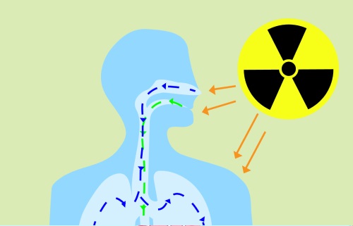
In medical imaging, radiation dose is usually reported in millisieverts (mSv). A millisievert is a way of describing how much ionizing radiation your body has absorbed and how biologically meaningful that exposure is. It is not the same as the amount of X-ray energy fired by the machine. Instead, it accounts for which tissues were exposed and how sensitive those tissues are.
A few rough comparisons help put the number into context:
- Natural background radiation from soil, building materials, food, and cosmic rays averages around 3 mSv per year for most people, varying with altitude and geography.
- A single chest X-ray delivers roughly 0.1 mSv, which works out to about ten days of background radiation.
- A mammogram is around 0.4 mSv.
- A head CT is in the range of 2 mSv.
- A chest or abdominal CT typically falls between 5 and 10 mSv, depending on the protocol and the patient.
These are typical ranges, not fixed numbers. Modern scanners and protocols can lower these doses considerably without losing diagnostic quality.
Which scans use radiation and which do not
Not every imaging test uses ionizing radiation. Knowing which is which often takes the anxiety out of the conversation:
- Ultrasound uses sound waves only. No ionizing radiation. This is why it is the first-line test for many abdominal, vascular, obstetric, and musculoskeletal questions.
- MRI uses a strong magnetic field and radio waves. No ionizing radiation. MRI is excellent for soft tissues such as the brain, spine, joints, and abdomen, and is often preferred when the question can be answered without X-rays.
- X-ray uses a small amount of ionizing radiation. Useful for bones, chest, and many quick triage questions.
- CT scan uses ionizing radiation, similar in nature to X-rays, to build detailed cross-sectional images. Higher dose than an X-ray but indispensable for many emergencies and complex diagnoses.
- Nuclear medicine and PET-CT use a small injected radioactive tracer plus, in the case of PET-CT, a low-dose CT component. Doses depend on the tracer and protocol.
When two tests can answer the same question, doctors usually prefer the one that uses no ionizing radiation. This matters most for younger patients and for repeated follow-up imaging.
When concern about radiation dose is warranted
For a single medically indicated scan in an adult, the dose is usually a minor consideration compared with the diagnostic value. Concern becomes more relevant in a few specific situations:
- Children and young adults have more years of life ahead during which any theoretical risk could matter, and their tissues are more radiation-sensitive than older adults'.
- Pregnancy: the fetus is more sensitive than mature tissues, especially in the first trimester. Non-ionizing options are preferred when they can answer the clinical question.
- Repeated imaging over months or years: patients with chronic conditions that require frequent CT (kidney stones, complex cancers, vascular surveillance) can accumulate meaningful cumulative doses. The strategy is usually to alternate modalities or use lower-dose protocols.
- Scans that may not change management: if a CT will not change what your doctor does next, it is reasonable to ask whether it is really needed. Most physicians welcome this question.
For one-off, clinically justified scans in healthy adults, the radiation conversation is usually a short one.
How radiology teams keep doses low
Modern radiology operates under the principle of ALARA, short for "as low as reasonably achievable." That principle is built into both the equipment and the workflow:
- Low-dose CT protocols have become standard for lung-cancer screening, kidney stones, and many other indications. They use a fraction of the dose of a routine CT while still answering the clinical question.
- Iterative reconstruction and AI-based denoising let scanners deliver less radiation while producing diagnostic-quality images.
- Automatic exposure control tailors the dose to the patient's size in real time.
- Pediatric-specific protocols follow Image Gently and similar campaigns, with dose adjusted for the child rather than borrowed from adult settings.
- Justification at the order step: radiologists and ordering physicians weigh whether ultrasound or MRI could answer the question first.
If you want to know the specifics for your scan, ask the technologist or radiologist. They are used to the question and can usually give you a ballpark dose in mSv.
Special populations: pregnancy and pediatrics
Two groups deserve a closer look. In pregnancy, ultrasound and MRI without contrast are the default when they can answer the clinical question. When an X-ray or CT is genuinely needed, abdominal shielding and reduced-dose protocols are used. The actual fetal dose from a single chest X-ray or a head CT is usually very small. Tell the imaging team before the scan if you are or might be pregnant.
In pediatrics, scanners are dialed down to match the child's size, and many questions that would be answered with CT in an adult are answered with ultrasound or MRI in a child. The American College of Radiology and the Image Gently Alliance publish protocols specifically for this. Children's hospitals routinely use them.
Why a second read can help
Once a scan has been done, the radiation is in the past. What matters next is whether the images are read carefully. A second radiology read can pick up findings that were missed, soften over-called findings, and answer the question of whether another scan is even needed. DocOrbit provides an expert second-opinion radiology report you can share with your own physician, which is especially useful before agreeing to a repeat CT or a more invasive next step. If your scan was related to a possible cancer, it is also worth reading how second opinions affect cancer outcomes.
Is the radiation from a CT scan dangerous?
For a single medically indicated CT scan in an adult, the radiation dose is small and the diagnostic benefit almost always outweighs the theoretical risk. The dose for a typical abdominal CT is in the range of a few millisieverts, which is comparable to a year or two of natural background radiation. Risk is cumulative, so the more relevant question is usually about repeated CTs over a lifetime, not a single scan.
Do MRI and ultrasound use radiation?
No. MRI uses a strong magnetic field and radio waves, and ultrasound uses sound waves. Neither involves ionizing radiation. That is one of the reasons doctors often choose MRI or ultrasound for younger patients, pregnant women, and follow-up imaging when either modality answers the clinical question.
Is it safe to have a scan during pregnancy?
Ultrasound and MRI without contrast are generally preferred during pregnancy because they do not use ionizing radiation. When an X-ray or CT is necessary, abdominal shielding and dose-reduction protocols are used, and the actual fetal dose is usually very low. Tell the imaging team if you are or might be pregnant before any scan so they can plan accordingly.
How much radiation is too much in a lifetime?
There is no precise threshold below which radiation is risk-free and above which it is dangerous. Regulators and radiology societies use the principle of keeping medical exposure as low as reasonably achievable while still answering the clinical question. For most patients, the cumulative dose from medically justified imaging over a lifetime is small compared with the diagnostic benefits.
Can children safely have CT scans?
Yes, when they are needed. Children are more radiation-sensitive than adults, so pediatric protocols use lower doses and limit scans to clear clinical indications. Many children's hospitals follow Image Gently guidelines, which adjust dose for the child's size and skip unnecessary phases. When ultrasound or MRI can answer the question, they are usually chosen first.
Key takeaways
- Ultrasound and MRI do not use ionizing radiation. X-rays and CT scans do, and of the two, CT carries the higher dose.
- A typical chest X-ray is around 0.1 mSv; a typical CT is in the range of a few to ten mSv, compared with about 3 mSv per year from natural background radiation.
- For a single medically indicated scan in an adult, the diagnostic benefit almost always outweighs the small theoretical risk.
- Children, pregnant patients, and those needing repeated imaging deserve extra attention to dose and modality choice.
- Modern dose-reduction techniques and second-read services can both reduce unnecessary exposure.
This article is for general information only and is not medical advice. Always discuss your imaging results and any next steps with a qualified physician.