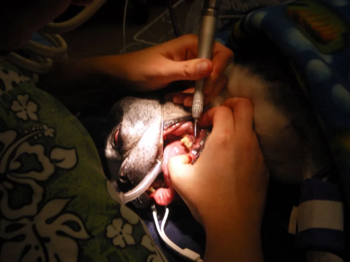
Most owners arrive at this diagnosis by one of two routes. Either the breath has been bad for so long that it became normal, or a firm swelling appeared under one eye overnight and looked like an insect bite. Then comes the part nobody expects: X-rays, a general anaesthetic, and a conversation about removing the tooth. The reassuring truth is that this is one of the most fixable problems in veterinary medicine. Dogs recover from dental surgery quickly, they eat well afterwards, and owners routinely describe a dog that seems years younger once the pain is gone.
What a tooth root abscess actually is
A tooth is not a solid block. Inside it sits the pulp, a living core of nerve and blood vessels. When bacteria reach that core, the pulp dies, and the infection drains out through the tip of the root into the bone of the jaw. That pocket of infection at the root tip is the abscess.
Bacteria get there by two main routes. The first is a fracture: a dog cracks a tooth on a bone, an antler, a stone, or a hard nylon chew, exposing the pulp directly. The upper fourth premolar, the large shearing tooth at the back of the upper jaw, is the classic casualty because it takes the most force. The second route is periodontal disease, where plaque hardens into tartar, inflames the gum, and slowly destroys the ligament and bone that hold the root in place until infection tracks down alongside it. The two often arrive together, and by the time a swelling appears, the process has usually been running for months.
Why the disease is invisible above the gumline
This is the single most useful thing to understand, because it explains everything else about how the problem is handled.
Around two thirds of a dog's tooth is buried: the root, the ligament that suspends it, and the bone socket around it. What you and your vet can see in a conscious mouth is the crown, the visible tip. Periodontal disease and root abscesses are diseases of the buried part. A crown can look clean and white while the bone underneath it has been eaten away, and a tooth that looks stained and unhappy can turn out to be perfectly well anchored.
Even the tartar you can see only tells you where plaque has been sitting, not how much attachment has been lost. A probe measures pocket depth but cannot see through bone. That is where imaging comes in.
The signs owners actually notice
Almost nobody brings a dog in suspecting a dead tooth. They come in for something else.
- Breath that has become sharply unpleasant rather than just doggy, often blamed on food or on the breed
- A firm swelling below one eye, sometimes bursting and draining a small amount of pus onto the cheek. This is the classic sign of an abscessed upper fourth premolar and is regularly mistaken for a bite, a wound, or a skin lump
- Chewing on one side only, dropping kibble, or picking food up and putting it down again
- Pawing at the face, rubbing the muzzle along the sofa, or shying away from being stroked on one side of the head
- Suddenly preferring soft food, or eating more slowly and less enthusiastically
- Drooling, sometimes tinged with blood, or a reluctance to play with toys that used to be favourites
- A quieter, flatter dog that owners describe afterwards as having been "just older"
The last one deserves emphasis. Dogs are extremely poor at showing dental pain. They keep eating, because not eating is not an option their instincts offer them. The change is usually in mood and energy rather than in appetite, which is exactly why it gets attributed to age.
Why full-mouth dental radiographs change the plan
Dental X-rays are not the same as the chest or abdominal films you may have seen before. Small sensors are placed inside the mouth against the teeth, and each area of the jaw is imaged separately, which is why a full-mouth series takes ten to fourteen individual views. What those images reveal changes the treatment decision constantly:
- Bone loss around the root that is invisible on examination, which decides whether a tooth can be saved or must come out
- A dark halo at the root tip, the radiographic signature of an abscess, confirming that the pulp is dead
- Root fractures and resorption, where the body is dissolving the root itself, a common and painful finding
- Retained root fragments from an old extraction or a tooth that broke off at the gumline years ago
- Unerupted teeth trapped in the bone, which can form cysts that quietly hollow out the jaw
- Bone changes that do not fit infection at all, which occasionally redirects the workup toward a jaw mass rather than a dental problem. Our article on bone tumours and osteosarcoma in dogs covers what that pattern looks like
Veterinary dental studies have repeatedly found that a substantial share of clinically important findings would have been missed without radiographs, in both directions: teeth that looked fine but needed removing, and teeth marked for extraction that were left in place once the images showed healthy bone.
Why it all has to happen under anaesthesia
This is where a lot of owners hesitate, and the hesitation is reasonable. The answer is practical rather than dramatic. A dental sensor has to sit still inside the mouth at a precise angle against the tooth being imaged, pockets have to be probed millimetre by millimetre, and tartar has to be removed from below the gumline, which is the only part that matters clinically. None of that is achievable in a conscious animal.
Anaesthesia also allows a protected airway, so water and bacteria from the cleaning cannot be inhaled, and proper pain relief including local nerve blocks. So-called anaesthesia-free dental cleaning polishes the visible crown and leaves the disease untouched, while giving the reassuring appearance that something has been done.
Before the day, expect a pre-anaesthetic assessment: examination, bloodwork, and attention to any existing condition. That matters more than age itself, and it is a familiar step for owners managing conditions such as chronic kidney disease in an older animal.
What treatment looks like
Extraction is the most common answer, and for a tooth with a dead pulp and lost bone support, it is a definitive one. Multi-rooted teeth are usually sectioned into pieces first so each root can be lifted cleanly, and the gum is closed with dissolving sutures.
Root canal treatment saves the tooth by cleaning out and sealing the dead pulp. It is a genuine option for strategically important teeth, such as the large canines or that upper fourth premolar, in a dog whose surrounding bone is still healthy. It is performed by veterinary dentists and needs radiographic follow-up.
Antibiotics alone are not a treatment. They will settle a facial swelling impressively, which is why the problem so often appears to resolve, but they cannot reach dead tissue inside a tooth. The infection returns, usually within weeks of the course finishing.
Recovery is generally faster than owners expect: soft food for one to two weeks, no hard chews or tug toys while the gum heals, prescribed pain relief as directed, and a recheck to confirm the sites have closed.
Preventing the next one
Daily toothbrushing with a pet-specific paste is the only measure that reliably slows plaque, and even a few days a week is meaningfully better than none. Dental diets, approved chews, and water additives help at the margins but do not replace brushing.
The point owners find most surprising is what to avoid: bones, antlers, hooves, ice cubes, and hard nylon toys are common causes of the fractures that start the whole process. If you cannot dent it with a thumbnail, it is hard enough to break a tooth.
Why a second read can help
Dental radiographs are technically demanding images. The angle at which the beam meets the root changes how the bone around it appears, and a shadow that suggests an abscess on one view can look entirely different on the next. Distinguishing early bone loss from normal anatomy, or an infected root tip from a more concerning bone pattern, takes practice with these specific views. DocOrbit can arrange an independent specialist review of your dog's dental radiographs and return a written report you can take to your own vet, which is often useful when several teeth are borderline or when the recommendation involves losing a strategically important tooth. The same reasoning applies whenever imaging drives an irreversible decision, as we discuss in our piece on Cushing's disease in dogs.
Can a tooth root abscess in a dog heal without extraction?
Antibiotics reduce the swelling and the pain, often within a couple of days, which is why the problem looks solved. They do not reach the dead tissue inside the tooth, so the infection returns once the course finishes. Lasting resolution means either removing the tooth or performing root canal treatment to sterilise and seal the inside of it. The choice between the two depends on the tooth, the amount of bone left around it, and what the animal uses that tooth for.
Why does my dog need X-rays for a tooth that looks fine?
Roughly two thirds of every tooth sits below the gumline, and that is where periodontal disease and root abscesses live. A crown can look clean and still sit on a root surrounded by destroyed bone. Studies of dogs having routine dental procedures have repeatedly found significant findings on X-ray in teeth that looked normal on examination, and normal-looking X-rays in teeth that had been marked for extraction. The images are what turn a guess into a plan.
Is dental anaesthesia safe for an older dog?
Age on its own is not a disease, and modern veterinary anaesthetic protocols are adjusted to the individual patient rather than to the birthday. What matters is the pre-anaesthetic assessment: bloodwork, heart and lung examination, blood pressure, and any medication the dog is already on. Many older dogs are anaesthetised safely every day for exactly this procedure. It is also worth remembering that leaving a painful infected tooth in place carries its own ongoing cost.
Will my dog be able to eat normally after teeth are removed?
Yes, and usually better than before. Dogs do not chew their food the way people do, and they cope remarkably well with missing teeth, including dogs that end up with very few left. Most owners report that appetite and enthusiasm at mealtimes improve once the source of pain is gone. Soft food is normally advised for the first week or two while the gum heals.
Is bad breath in dogs a sign of dental disease?
Most of the time, yes. Healthy dog breath is not pleasant but it is fairly neutral, whereas the sharp, rotten smell owners describe usually comes from bacteria under the gumline. Bad breath is one of the earliest and most reliable signals that periodontal disease is present. It is worth mentioning at a routine appointment rather than accepting it as normal for the breed or the age.
Key takeaways
- A tooth root abscess is infection at the tip of a root, usually following a fractured tooth or advanced periodontal disease
- A firm swelling under the eye is the classic sign and is frequently mistaken for an insect bite or a wound
- Two thirds of every tooth is below the gumline, so the disease is invisible without radiographs
- Full-mouth dental X-rays regularly change which teeth are removed and which are saved
- Antibiotics settle the swelling but do not cure it; extraction or root canal treatment does
- Dogs eat well after extractions, and most owners notice a brighter, more comfortable animal within weeks
How do I have my pet's X-ray reviewed by another veterinary radiologist online?
Yes. Online veterinary radiology second opinions run entirely from home: you upload your images, with no CD to post and no second trip, and a subspecialist veterinary radiologist re-reads the study and writes a structured report you can give to your own vet. DocOrbit returns that signed second read in 24 to 48 hours. You can then message the veterinary radiologist with questions about the X-ray and about what the report actually means for your pet.
This article is for general information only and is not veterinary advice. Always discuss your animal's imaging results and next steps with a qualified veterinarian.
