If you have just been told your dog has a liver shunt, the first feeling is often not fear but recognition: all those months of the puppy who stayed small, who went quiet and strange after dinner, who took forever to wake up after the neutering anaesthetic, suddenly line up into one explanation. The reassuring part is that this is a plumbing problem more than a disease of the liver itself, it is often correctable, and a large proportion of dogs with the common type go on to a normal life after treatment. Here is what a shunt actually is, why it hides for so long, and what each test is really doing.
What a liver shunt actually is
Blood leaving the stomach and intestines is full of things absorbed from food, including ammonia and other by-products of protein digestion. That blood is supposed to travel through the portal vein straight into the liver, which filters it before it reaches the rest of the body. The liver is the customs checkpoint.
A portosystemic shunt is an abnormal blood vessel that bypasses that checkpoint. Unfiltered blood goes straight into the general circulation, and the toxins it carries reach the brain. At the same time, the liver is starved of the growth factors that arrive in portal blood, so it stays small and underdeveloped.
That single anatomical fact explains almost every sign these dogs show.
The three types, and why the label matters
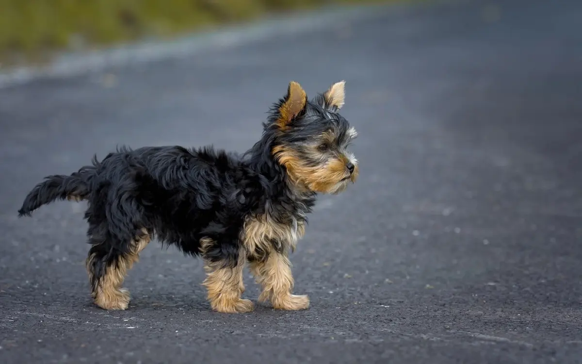
- Congenital extrahepatic — a single abnormal vessel outside the liver, present from birth. This is the most common form and is typical of small breeds: Yorkshire Terriers, Maltese, Shih Tzus, Pugs, Miniature Schnauzers, Havanese, Jack Russells. It is also the form most likely to be fixed surgically
- Congenital intrahepatic — the vessel runs inside the liver, usually a fetal channel that never closed after birth. More typical of large breeds such as Irish Wolfhounds, Labradors, Golden Retrievers, and Australian Shepherds. Harder to reach surgically, so interventional or medical approaches are used more often
- Acquired (multiple) — many small shunts that open up later in life because pressure in the portal system has risen, usually because of existing liver disease such as cirrhosis. These are the body compensating, not a birth defect, and they are not closed surgically. Treatment targets the underlying liver disease
When a vet says the type matters, this is why: the same test results can lead to a curative operation in one dog and lifelong medical care in another.
Why it goes unnoticed for months
Almost every owner of a shunt dog has been told at some point that the puppy is just small, or just a fussy eater, or just sleepy. That is not negligence, it is genuinely how the condition presents. The signs are intermittent, they come and go with meals, and they are easy to attribute to a young animal's personality.
The classic picture looks like this:
- A puppy who is noticeably smaller and lighter than littermates and does not catch up
- Periods of being dull, dopey, disoriented, wobbly, or head-pressing against a wall, typically starting one to three hours after eating and then resolving
- Poor or picky appetite, sometimes vomiting or intermittent diarrhoea
- Drinking and urinating more than expected for the size of the dog
- Seizures in a puppy with no other explanation
- Excessive drooling, which is more typical of cats but does occur in dogs
- Blood or straining in the urine, from ammonium biurate bladder stones that form because of the same metabolic problem
- An unusually slow, prolonged recovery from anaesthesia or sedation, because the liver cannot clear the drugs at a normal rate
That last one is worth dwelling on, because it is how a large number of shunts are finally caught. A routine spay or neuter turns into a dog who will not wake up properly, and the practice starts looking at liver function.
If your dog's main presenting sign was seizures, it is also worth understanding how the neurological workup separates a metabolic cause like this from a structural one; we cover that in our article on seizures and brain tumours in dogs.
What the bloodwork shows
Routine bloodwork rarely gives a clean answer, but it leaves a very characteristic set of fingerprints.
- Bile acid stimulation test — the single most useful screening test. A sample is taken after fasting, the dog is fed a small fatty meal, and a second sample is taken two hours later. In a shunt dog the post-meal value is dramatically high, because bile acids that should be recycled by the liver spill into the general circulation instead
- Low blood urea nitrogen — counterintuitive, but the liver is where ammonia is converted into urea. A liver that is bypassed and underdeveloped makes less urea, so a low value in a young small-breed dog is a genuine flag
- Low albumin, low cholesterol, low blood glucose — all products of a liver that is not doing its full job
- Microcytic anaemia — red cells that are smaller than normal, related to disturbed iron handling. Often mild and easy to overlook on a report
- Raised blood ammonia — supports the diagnosis, though the sample handling is fussy and a normal value does not exclude a shunt
- Urine — ammonium biurate crystals or stones are a strong pointer
Bile acids plus a low urea in a young small-breed dog is the combination that usually triggers the referral conversation. Liver enzymes, by contrast, are often only mildly raised or even normal, which is another reason the diagnosis slips through.
What imaging shows, and why ultrasound alone can miss it
Bloodwork tells you the liver is being bypassed. Imaging has a harder job: it has to find the actual vessel, and show the surgeon exactly where it runs, where it drains, and what surrounds it.
Ultrasound is the usual first step, and it is genuinely useful. It shows a small liver, often kidneys that look large for the dog, sometimes bladder stones, and with a skilled operator and a cooperative patient it can visualise the shunting vessel itself and the direction of flow on Doppler. It is non-invasive, needs no general anaesthetic, and costs a fraction of the alternative.
But a single small abnormal vessel among a network of normal ones is a hard target. Bowel gas obscures the view, deep-chested and larger dogs are harder to scan, intrahepatic shunts hide among normal liver vasculature, and results vary considerably with operator experience. A normal-looking ultrasound in a dog with clearly abnormal bile acids does not close the case; it usually means the next test is needed.
CT angiography is now the reference standard. Contrast is injected and a rapid scan captures the vascular anatomy in three dimensions. It reliably distinguishes a single shunt from multiple ones, shows extrahepatic from intrahepatic, and gives the surgeon a road map before they open the abdomen. It needs a short general anaesthetic and a referral centre, which is where the cost and the anxiety come in for most owners, but it is what turns maybe into a plan.
The honest way to think about the sequence is that ultrasound is a good screening and problem-finding tool, and CT is a surgical planning tool. Vets are not being thorough for the sake of it when they recommend the second one after the first.
Treatment: medical management and surgery
Medical management is almost always started first, whether or not surgery is planned, because it stabilises the dog and makes anaesthesia safer.
- Diet — moderate, high-quality protein rather than severe restriction. These dogs still need protein to grow. Prescription hepatic diets are built for this, and dairy or vegetable protein sources are generally handled better than red meat. Several small meals rather than one large one smooths out the after-meal signs
- Lactulose — a syrup that acidifies the colon so ammonia is trapped there and passed out rather than absorbed. The dose is titrated to give soft but not liquid stools
- Antibiotics — usually metronidazole or amoxicillin, to reduce the gut bacteria that produce ammonia in the first place
- Anti-seizure medication — used in selected cases, particularly around the time of surgery in higher-risk dogs
Medical management controls signs well in many dogs, but it does not fix the anatomy, and the liver continues to receive less blood than it needs.
Surgical attenuation aims to close the abnormal vessel and redirect blood back through the liver. This is almost never done by tying it off in one go, because the liver's own vasculature is underdeveloped and cannot absorb the full flow immediately. Instead, a device is placed that narrows the vessel gradually over weeks: an ameroid constrictor, a cellophane band, or a thin film band. For intrahepatic shunts, an interventional radiology approach placing a stent and coils through a vein is sometimes possible without opening the abdomen.
Recovery involves several weeks of confinement, continued diet and medication, and a repeat bile acid test a few months later to see whether treatment can be withdrawn. The main serious complication to know about is post-operative seizures, which are uncommon but are the reason surgeons monitor these dogs closely in the days after the operation.
Prognosis, and the guilt question
For a single congenital extrahepatic shunt closed with a gradual-attenuation device, outcomes are good. The large majority of dogs improve substantially, most eventually come off medication and special diet, and many go on to a normal life expectancy. This is one of the more satisfying conditions in small animal practice precisely because a structural fix exists.
Intrahepatic shunts carry a somewhat higher risk and a more variable outcome, though results have improved with interventional techniques. Multiple acquired shunts are a different situation entirely, because the shunts are a consequence of liver disease rather than the primary problem, and the outlook follows the underlying disease.
On the guilt: dogs with shunts are routinely diagnosed at a year old, at three years old, sometimes later. The signs are intermittent, they mimic ordinary puppy behaviour, standard liver enzymes are often unremarkable, and the specific test that reveals it is one nobody runs without a reason to. Delay is the norm for this condition, not a failure of care. What matters is what happens now, and dogs diagnosed later still respond to treatment.
Why a second read can help
This is a diagnosis where the imaging carries an unusual amount of weight. Whether the shunt is single or multiple, extrahepatic or intrahepatic, and exactly where it drains determines whether your dog is a surgical candidate at all, and a hesitant or equivocal report can leave an owner stuck between an expensive referral and doing nothing. If you are holding an ultrasound or CT study and the conclusion feels uncertain, or the recommendation does not match how your dog seems day to day, DocOrbit can arrange an independent specialist review of the images and return a written report you can take to your own vet. It is the same reasoning we apply to other conditions where the imaging call directly decides the treatment path, such as Cushing's disease in dogs or a gallbladder mucocele.
Can a dog live a normal life with a liver shunt?
Many dogs do, particularly those with a single congenital shunt outside the liver that is closed surgically. A large majority of those dogs come off medication and diet restrictions within months and go on to a normal life expectancy. Dogs managed medically rather than surgically, and dogs with multiple acquired shunts, usually need lifelong diet and medication, and their outlook depends heavily on the underlying liver disease.
Can a liver shunt be missed on ultrasound?
Yes, and this is one of the most common reasons a diagnosis is delayed. A shunt is a single small abnormal vessel hidden among many normal ones, and finding it depends on the vessel's location, the dog's size, gas in the bowel, and the experience of the person scanning. A normal ultrasound in a dog with high bile acids does not rule a shunt out, which is why CT angiography is usually the next step rather than the end of the search.
Is liver shunt surgery in dogs worth it?
For a single congenital extrahepatic shunt, surgery generally offers the best long-term outcome, and most dogs that recover well from the procedure need little or no ongoing treatment afterwards. It is real surgery with real risk, including a small chance of post-operative seizures, so it is a decision made with a surgeon who has assessed your individual dog. Medical management is a legitimate choice, not a failure, when surgery is not possible or not affordable.
What are the first signs of a liver shunt in a puppy?
The earliest signs are usually being smaller and thinner than littermates, a poor appetite or fussy eating, and periods of being quiet, wobbly, or vacant that come on an hour or two after a meal and then pass. Drinking and urinating more than expected is common. Many owners also notice their puppy takes an unusually long time to wake up from a routine anaesthetic, which is often the clue that leads to testing.
What should a dog with a liver shunt eat?
The usual approach is a diet with moderate, high-quality protein rather than a low-protein one, because these dogs still need protein to grow and maintain muscle. Prescription hepatic diets are formulated for exactly this balance, and dairy or plant-based protein sources are often better tolerated than red meat. Feeding several small meals a day rather than one large one helps limit the after-meal wobbliness, and your vet will tailor the plan to your dog.
Key takeaways
- A shunt is an abnormal vessel that lets gut blood bypass the liver, which explains the small size, the after-meal dullness, and the slow anaesthetic recovery
- High post-meal bile acids together with a low urea in a young small-breed dog is the combination that should prompt a shunt workup
- Ultrasound is a good first look but genuinely can miss a shunt; a normal scan does not close the question
- CT angiography defines the anatomy and is what a surgeon needs to plan attenuation
- Single extrahepatic shunts closed with a gradual-attenuation device have good outcomes, and many dogs eventually need no treatment at all
- Late diagnosis is the norm with this condition; dogs found at one, two, or three years old still do well
How do I have my pet's scan reviewed by another veterinary radiologist online?
Yes. Online veterinary radiology second opinions run entirely from home: you upload your images, with no CD to post and no second trip, and a subspecialist veterinary radiologist re-reads the study and writes a structured report you can give to your own vet. DocOrbit returns that signed second read in 24 to 48 hours. You can then message the veterinary radiologist with questions about the scan and about what the report actually means for your pet.
This article is for general information only and is not veterinary advice. Always discuss your animal's imaging results and next steps with a qualified veterinarian.
