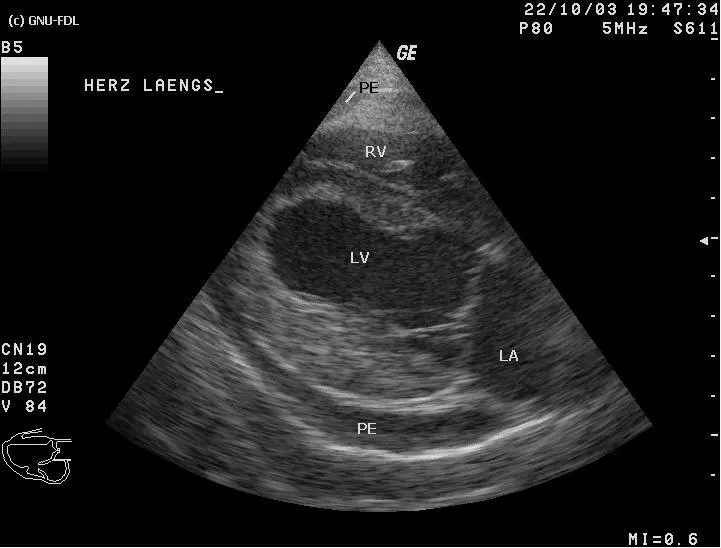
If your dog collapsed, seemed suddenly weak, or had a swollen belly, and the echocardiogram report used the words pericardial effusion or fluid around the heart, it is normal to feel frightened. Here is the reassuring part: the fluid itself is usually the easiest part of this problem to fix, and dogs often perk up within minutes of it being drained. What takes more time, and often benefits from a specialist's eye, is figuring out why the fluid built up in the first place. This article covers what pericardial effusion actually is, why it becomes a true emergency, what usually causes it, and what the days after the emergency drain typically look like.
What is pericardial effusion?
The pericardium is a thin, double-layered sac that wraps around the heart and normally holds only a few drops of fluid, just enough to let the heart beat inside it without friction. Pericardial effusion is an abnormal buildup of fluid between those two layers, and on an echocardiogram it shows up as a dark, fluid-filled space outlining the heart, sometimes with a speckled texture if the fluid is bloody rather than clear.
- The pericardium normally holds only a thin lubricating film of fluid, not a measurable pocket
- Pericardial effusion is an abnormal buildup of fluid between the two layers of that sac
- On echocardiogram it appears as a dark space surrounding the heart, and it can look speckled rather than plain black when the fluid is blood
- In a large effusion, the heart can appear to swing within the fluid with every beat, a finding vets call a swinging heart
Why it is a true emergency: cardiac tamponade
As fluid builds up, pressure inside the pericardial sac rises. The heart's right-side chambers are thin-walled and normally work at low pressure, so they are the first to be squeezed shut between beats, long before the fluid rises high enough to affect the thicker-walled left side. That squeeze stops the heart from filling properly, so less blood goes out with each beat no matter how hard the heart tries, a state called cardiac tamponade. The result is the kind of emergency described for a bloated, twisted stomach in dogs: a problem that gets worse by the hour rather than something you can watch overnight. A dog in tamponade needs to be seen the same day, not the next morning.
What causes fluid around the heart
Once tamponade is relieved, the real question becomes why the fluid appeared. The common causes are:
- Cancer, most often hemangiosarcoma growing from the right atrium or its small pouch-like auricle, the single most common cause found in middle-aged to older, large-breed dogs
- A heart base tumor, also called a chemodectoma or aortic body tumor, which tends to grow slowly and is more typical in Boxers, Bulldogs, and other short-nosed breeds
- Idiopathic (benign) hemorrhagic pericarditis, where no cause is found even after a full workup, notably common in Golden Retrievers
- A tear in the wall of the left atrium from severe, long-standing mitral valve disease, which lets blood leak directly into the pericardial sac
- Less commonly, infection, a migrating foreign body, or a bleeding disorder such as rodenticide (rat poison) toxicity
The warning signs to know
- Sudden weakness or collapse — sometimes right after exercise or excitement.
- Pale, grey, or bluish gums.
- A swollen belly — as pressure backs up and fluid leaks into the abdomen, a distension that can look similar to what shows up with a bleeding splenic mass on ultrasound, since the same tumor can sometimes cause both.
- Rapid, shallow breathing and weak pulses.
- Restlessness, or reluctance to lie down comfortably.
You do not need to see all of these. Sudden collapse or weakness together with a swollen belly is enough to go straight to a veterinarian.
How the echocardiogram diagnoses it
Echocardiography is the definitive test: it shows the fluid directly, measures how much has collected, and can look for a mass at the heart base or right atrium that might explain it. A chest X-ray usually shows a rounded, oversized heart shadow, and an ECG can show small, alternating-height beats called electrical alternans, but neither can see the fluid or a mass as clearly as the echo does. Finding the fluid is the easy part of the scan. Finding, or ruling out, a small mass at the heart base or right atrium is much harder, especially in an unstable dog scanned quickly in an emergency room, and it is exactly the kind of detail a second, unhurried look at the same images can catch.
Emergency treatment: pericardiocentesis
The immediate treatment is pericardiocentesis: a needle or small catheter is passed into the pericardial sac, usually with ultrasound guidance and the heart rhythm monitored throughout, and the fluid is drained. Relief is often dramatic, with dogs breathing easier and standing up within minutes. A sample of the fluid may be sent for analysis, though blood-tinged effusions are notoriously hard to read under the microscope, since the blood itself can hide the cancer cells a pathologist is looking for.
Finding the cause and what comes next
Once your dog is stable, the workup turns to why the fluid appeared. That usually means a more careful echocardiogram, sometimes alongside a CT scan or an abdominal ultrasound to check the spleen and liver for spread, since hemangiosarcoma is well known for turning up in more than one organ at once. If a resectable mass is found, surgery may be offered. Even when the mass cannot be fully removed, as is often the case with a heart base tumor, a pericardiectomy, removing a window of the pericardial sac, stops fluid from collecting around the heart again and can give many dogs a good quality of life for a long time. For hemangiosarcoma the outlook stays more guarded because it tends to have already spread by the time it is found, but draining the fluid and, when appropriate, surgery can still buy meaningful comfortable time with your dog.
Why a second read can help
So much of this hinges on a small structure that is easy to miss on a first look: a heart base or right atrial mass a few millimeters across, seen on a scan performed while a dog was unstable and time was short. If your dog's echo report leaves the cause of the effusion uncertain, or a mass is described as possible rather than definite, asking for an online veterinary radiology second opinion is a reasonable next step. A subspecialist reviews the same images away from the pressure of an emergency room and either confirms what was seen or characterizes a finding the first pass did not have time to pin down. Many owners choose to pair that read with the advice of an online veterinary radiologist before deciding on surgery, so the decision rests on the clearest possible picture of your dog's heart.
Is pericardial effusion in dogs always caused by cancer?
No. Cancer, especially a tumor called hemangiosarcoma on the right side of the heart, is the single most common cause found in dogs with pericardial effusion, but it is not the only one. A meaningful share of cases, particularly in Golden Retrievers, turn out to be idiopathic, meaning no cause is found even after a full workup, and these dogs often do well long-term. A heart base tumor or a tear related to long-standing valve disease are less common causes. Imaging, and sometimes fluid analysis, is the only way to know which applies to your dog.
Which dog breeds are most at risk of pericardial effusion?
German Shepherds and Golden Retrievers are the breeds diagnosed most often overall, though for different reasons: Golden Retrievers are strongly linked to the idiopathic benign form, while German Shepherds and other large breeds are overrepresented among dogs with hemangiosarcoma. Boxers, Bulldogs, and Boston Terriers are the classic breeds for heart base tumors. That said, any dog of any breed can develop pericardial effusion.
Will the fluid come back after it is drained?
It depends on the cause. Idiopathic effusion recurs in some dogs, but many never have a second episode after the first drain. Effusion caused by a tumor tends to come back much sooner, sometimes within weeks, which is why dogs with a tumor-related effusion are often offered a pericardiectomy, a surgery that removes part of the pericardial sac so fluid can no longer collect around the heart even if the underlying mass is still there.
How long can a dog live with pericardial effusion?
It varies enormously and depends almost entirely on the cause. Dogs with idiopathic pericardial effusion often go on to live a normal lifespan. Dogs with a heart base tumor, which tends to grow slowly, can do well for a long time, especially after a pericardiectomy. Hemangiosarcoma carries a more guarded outlook because it has usually already spread by the time it is found, though draining the fluid and surgery can still buy meaningful comfortable time. Your vet or a cardiologist can give you a realistic range once the cause is known.
Key takeaways
- Pericardial effusion is fluid that builds up around the heart and can compress it, causing a life-threatening drop in blood flow called cardiac tamponade.
- Sudden weakness or collapse, pale or grey gums, and a swollen belly, especially together, mean same-day veterinary care.
- Draining the fluid (pericardiocentesis) relieves the emergency almost immediately; finding the underlying cause takes imaging and sometimes more time.
- Hemangiosarcoma, heart base tumors, idiopathic pericarditis, and a valve-disease-related tear are the main causes, and the outlook differs hugely between them.
- Fluid that keeps coming back, whatever the cause, is often managed with a pericardiectomy so it can no longer collect around the heart.
Can I get a second opinion on my dog's echocardiogram online?
Yes. Online veterinary radiology second opinions run entirely from home: you upload your images, with no CD to post and no second trip, and a subspecialist veterinary radiologist re-reads the study and writes a structured report you can give to your own vet. DocOrbit returns that signed second read in 24 to 48 hours. You can then message the veterinary radiologist with questions about the echocardiogram and about what the report actually means for your dog.
This article is for general information only and is not veterinary advice. Always discuss your animal's imaging results and next steps with a qualified veterinarian.
