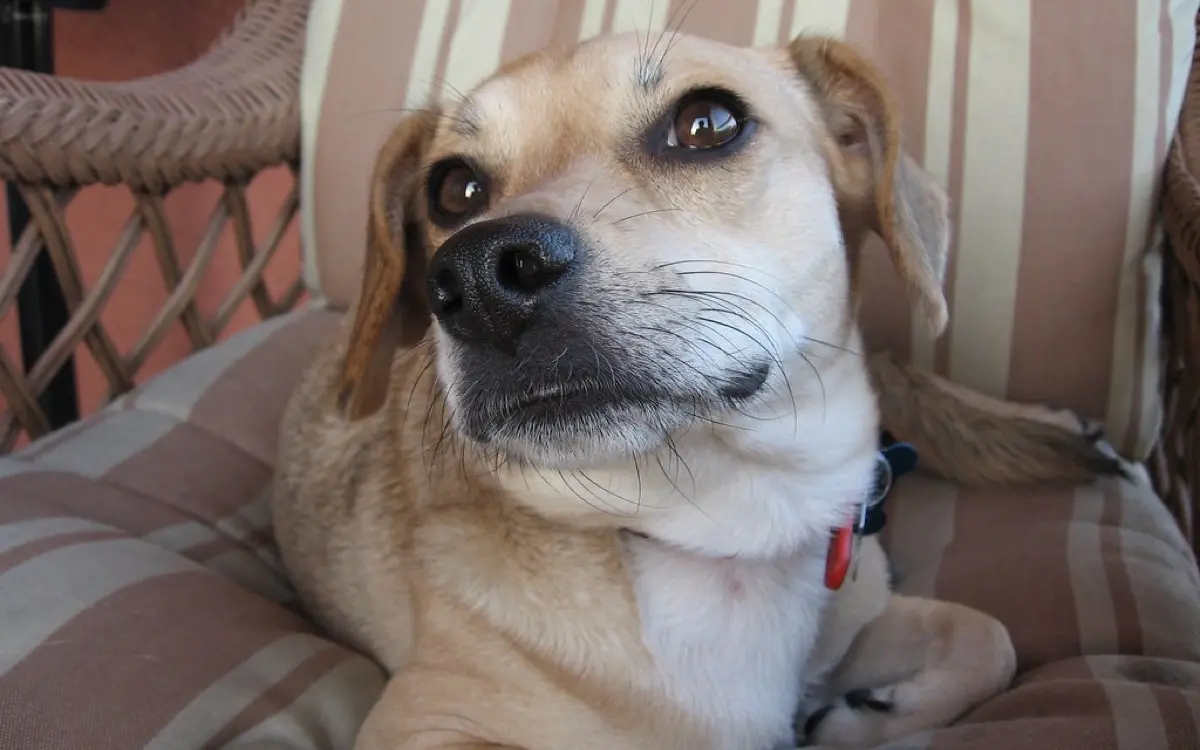
It started as the occasional sneeze. Now you are wiping the same nostril several times a day, there is a crust on the cushion in the morning, and you are on the third course of antibiotics that helped for a week and then stopped helping. Two things are worth knowing before you read any further. First, a long-running nose problem in a dog almost always has a specific physical cause sitting inside the nose, which means there is usually something to find. Second, the single most useful clue costs nothing: whether the discharge comes from one nostril or both.
Chronic nasal disease is one of the most under-investigated problems in general practice, mostly because the nose is genuinely difficult to see into. Repeated antibiotic courses without a diagnosis are the usual pattern, and they rarely solve anything on their own.
What the discharge itself tells you
Before any imaging, your vet is reading a few details that narrow the list considerably.
- One nostril or both. One side points to something physically located in that side of the nose. Both sides suggests something affecting the whole airway or the whole body, or a one-sided problem that has since crossed the midline.
- What it looks like. Clear and watery, thick and mucoid, yellow-green, or blood-tinged. Discharge that changes over weeks from clear to thick to bloody is a progression worth mentioning.
- How it started. A sudden fit of violent sneezing during or right after a walk in long grass is the classic story for an inhaled grass seed. A slow, creeping onset over months is a different story entirely.
- Airflow. Hold a small mirror or a wisp of fur near each nostril. Reduced airflow on one side is a genuinely useful observation, and it is one owners can make at home.
- The face itself. Any swelling over the bridge of the nose, a change in the shape of the muzzle, an eye that looks pushed forward, or a nostril that has lost its normal black pigment and looks ulcerated.
One thing that alarms owners and usually should not: reverse sneezing, the sudden episode of loud snorting inward that looks like the dog cannot breathe. It typically lasts under a minute, resolves on its own and is not the same as chronic nasal disease.
The common causes
Most chronic nasal cases in dogs come down to a short list.
- A foreign body, usually a grass seed or awn. Dramatic sneezing at onset, one-sided discharge afterwards, and completely curable once it is removed.
- Dental disease. The roots of the upper canine and the large upper cheek teeth lie directly against the nasal cavity. An abscess can erode through and drain into the nose, which is why our article on tooth root abscesses on dental X-ray is relevant far more often than owners expect.
- Fungal rhinitis, most often Aspergillus. It typically affects young to middle-aged dogs with longer noses, produces copious discharge, and often causes a painful nose with ulceration or loss of pigment at the nostril.
- Nasal tumours, usually in older dogs and more common in long-nosed breeds. The hallmark is slow, relentless progression, discharge that starts on one side, and intermittent bleeding.
- Chronic inflammatory rhinitis, a diagnosis reached once the others have been excluded. It is frustrating to manage but not dangerous.
- Bleeding and blood pressure problems, which cause nosebleeds without any disease inside the nose at all. These usually bleed from both nostrils and often come with bruising elsewhere.
Why plain X-rays are not enough
The inside of a dog's nose is filled with delicate scrolls of bone called turbinates, layered like the pages of a book. On a flat X-ray, those layers superimpose on one another and produce a grey blur that hides the very thing you are looking for. Even with a perfectly positioned skull radiograph under anaesthesia, subtle turbinate destruction is easy to miss.
A CT scan takes the nose apart into thin slices. On those slices a radiologist can assess which turbinates have been destroyed and on which side, whether the bone of the nasal septum, the hard palate or the orbit is intact, whether the frontal sinus is involved, and whether disease has reached the thin plate of bone that separates the nose from the brain. It also shows where the abnormal tissue actually is, which turns rhinoscopy from a blind search into a targeted one.
Patterns overlap more than textbooks suggest, but broadly: fungal disease tends to destroy turbinates and leave cavities with a thickened lining, sometimes with the nasal bones themselves looking thinned from the inside. Tumours tend to produce a soft-tissue mass that fills one side, destroys the bone around it and pushes across the midline or towards the eye. A foreign body may show as a small dense object, or only as a localised patch of inflammation around it.
What the workup usually looks like
Most dogs go under a single anaesthetic and come out with an answer. The usual sequence is a CT scan first, then rhinoscopy with a small camera, then biopsies of anything abnormal, plus dental X-rays if a tooth is under suspicion. Doing the CT first matters, because the scope and the biopsy forceps cause bleeding and swelling that would obscure the images if the order were reversed.
Bloodwork and a blood pressure check usually come first for a dog bleeding from both nostrils, and a chest X-ray is often added when a tumour is on the list.
Signs that warrant an earlier appointment
- Repeated bleeding from the same nostril over weeks
- Any change in the shape of the face or a firm swelling over the muzzle
- An eye that appears to bulge or sits differently from the other
- Complete loss of airflow through one nostril
- Loss of pigment, crusting or ulceration at the edge of the nostril
- A seizure or a sudden change in behaviour in a dog with known nasal disease, since it can mean disease has reached the front of the brain. Our piece on seizures and brain tumours in dogs on MRI covers what that assessment involves
- Loss of appetite or weight in combination with any of the above
Treatment, in plain terms
The good news is that several of the common causes are fixable. A grass seed is removed at rhinoscopy and the dog is usually normal within days. A diseased tooth is extracted and the discharge resolves. Fungal rhinitis is treated by flushing an antifungal solution through the nasal cavity under anaesthesia, and most dogs respond well, sometimes needing a second treatment.
Nasal tumours behave differently from many cancers because they stay local for a long time rather than spreading early to distant organs. Radiation therapy is the main treatment and often gives good control of the sneezing, discharge and bleeding along with a meaningful stretch of comfortable time, frequently many months to over a year depending on the tumour type and how far it has spread locally. Surgery on its own is rarely sufficient. Tumours in bone elsewhere in the body follow different rules, as described in our article on bone tumours on limb X-rays.
Chronic inflammatory rhinitis is managed rather than cured, with anti-inflammatory medication, flushing and humidified air, and periods of improvement between flare-ups.
Why a second read can help
Nasal CT is one of the harder reads in veterinary imaging. Fungal disease and tumour can produce overlapping appearances, early turbinate loss is subtle, and the assessment of whether the cribriform plate is intact often decides whether a dog is a candidate for a nasal flush at all. Those calls change the plan and the cost of what comes next. If your dog's scan report leaves your vet weighing two very different possibilities, an independent radiology opinion is a sensible step. DocOrbit lets you or your veterinary practice upload the images and get an expert second read to bring back into the conversation with your own team.
Is nasal discharge from one nostril worse than from both?
Not worse, but more informative. Discharge from a single nostril points towards something sitting inside one side of the nose, such as a grass seed, a diseased tooth root, a fungal infection or a growth. Discharge from both nostrils more often reflects something affecting the whole airway or the body as a whole, including infection, allergy or a clotting problem. Many one-sided problems eventually become two-sided as they progress, so the pattern at the start matters more than the pattern after months.
Why does my dog need a CT scan instead of an X-ray of the nose?
The nose is packed with paper-thin scrolls of bone, and on a flat X-ray they all overlap into a single grey haze that hides exactly what matters. A CT separates the nose into thin slices, so the radiologist can see which turbinates have been destroyed, whether the bone around them is intact, whether the frontal sinus is involved and whether anything has pushed towards the eye or the brain. That level of detail is what makes the difference between guessing and planning.
Can a bad tooth cause a runny nose in a dog?
Yes, and it is missed surprisingly often. The roots of the upper canine and the large upper cheek teeth sit directly against the nasal cavity. When one of them abscesses, infection can break through that thin partition and drain into the nose, producing discharge or sneezing on that side. Dental X-rays taken under anaesthesia are the only reliable way to see it, since the affected tooth frequently looks normal from the outside.
Is a nosebleed in a dog always a tumour?
No. A single nosebleed after rough play or a sneeze that irritates the lining is usually nothing. Repeated bleeding from the same nostril over weeks is the pattern that warrants investigation, and even then the causes include fungal infection, a foreign body and a clotting problem as well as a growth. Bleeding from both nostrils at once, or bruising elsewhere on the body, shifts the suspicion towards a blood-clotting or blood-pressure problem and makes bloodwork the first step.
Does my dog need anaesthesia for a nose CT?
Almost always, because the images must be perfectly still and precisely positioned to be readable. The upside is that vets usually plan everything into a single anaesthetic: the CT first, then rhinoscopy to look inside the nose with a camera, then biopsies of anything suspicious. Doing it that way means one anaesthetic instead of three, and the scan guides the camera to the right place rather than sampling blindly.
Are nasal tumours in dogs treatable?
They are treatable, and they behave differently from many other cancers because they tend to stay local for a long time rather than spreading early to other organs. Radiation therapy is the mainstay and often gives good symptom control along with a meaningful extension of comfortable life, frequently measured in many months to more than a year. Surgery alone is rarely enough. Outcomes vary considerably with tumour type and how far the disease has extended, so the CT and the biopsy together shape the conversation.
Key takeaways
- Weeks of sneezing or discharge in a dog almost always has a specific cause inside the nose, and repeated antibiotic courses rarely resolve it.
- One nostril versus both is the most useful early clue, and reduced airflow on one side is something you can check at home.
- Plain X-rays of the nose blur the turbinates together; CT is the imaging test that actually answers the question.
- The workup is usually a single anaesthetic combining CT, rhinoscopy and biopsy, in that order.
- Foreign bodies, tooth root abscesses and fungal infection are all treatable, and even nasal tumours respond well enough to radiation to be worth discussing properly.
Can I ask a veterinary radiologist about my pet's CT report online?
Yes. Online veterinary radiology second opinions run entirely from home: you upload your images, with no CD to post and no second trip, and a subspecialist veterinary radiologist re-reads the study and writes a structured report you can give to your own vet. DocOrbit returns that signed second read in 24 to 48 hours. You can then message the veterinary radiologist with questions about the CT and about what the report actually means for your pet.
This article is for general information only and is not veterinary advice. Always discuss your animal's imaging results and next steps with a qualified veterinarian.
