If your dog keeps bringing food back up and the chest X-ray report says "megaesophagus," the word sounds worse than the day-to-day reality. It means the esophagus, the muscular tube that carries food and water from the mouth to the stomach, has widened and lost much of its ability to push food along. Many dogs live comfortably for years once feeding is adjusted at home. The part that deserves real attention is aspiration, food or water slipping into the lungs, because that can cause pneumonia. Here is what the X-ray shows, what causes it, and how it is managed.
What megaesophagus is
A healthy esophagus is not a passive pipe. Its walls squeeze in coordinated waves, called peristalsis, that carry each swallow down to the stomach. In dogs it is built mostly of skeletal muscle steered by nerves, so nerve and muscle diseases hit it hard. In megaesophagus the wall weakens and stretches into a floppy, widened sac, and food and water sit there instead of moving on. "Mega" simply means large. Sooner or later the pooled contents slide back up, which is regurgitation.
There are two broad forms. In the congenital form a puppy is born with the problem and starts regurgitating when it moves onto solid food. The acquired form appears later, most often in middle-aged or older dogs. Because less food reaches the stomach, many affected dogs stay hungry and lose weight.
Regurgitation or vomiting: how to tell
The two can look alike on the kitchen floor, but they are different events, and describing which one you saw is one of the most useful things you can tell your vet.
- Regurgitation is passive. Food comes up with little or no warning and no heaving, is often undigested, and may be shaped like a tube coated in slimy mucus. It can happen minutes or hours after eating, and many dogs eat it again straight away.
- Vomiting is active. Nausea comes first, with drooling, lip licking and restlessness, followed by forceful belly contractions. The contents are partly digested and may be yellow with bile.
Some dogs do both, and even vets cannot always tell from the story alone, so a short phone video of an episode is worth bringing along.
What megaesophagus looks like on an X-ray
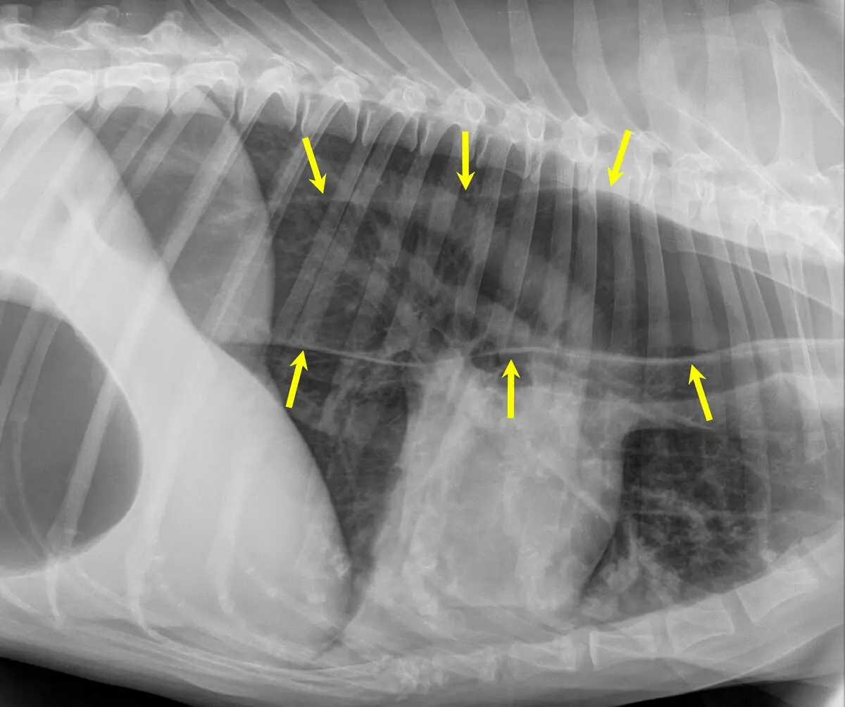
A normal esophagus is collapsed and usually invisible on a standard chest X-ray, because it has the same density as the tissue around it. In megaesophagus it fills with air, fluid or food, and shows up. Radiologists look for:
- A widened esophagus, a broad tube running through the chest above the heart and windpipe, sometimes along its whole length and sometimes in one stretch
- A "tracheal stripe," a thin white line where the wall of the air-filled esophagus lies against the back wall of the windpipe
- The windpipe, and sometimes the heart, pushed downward by the swollen esophagus
- Food or fluid inside, which looks like a patchy soft-tissue shadow rather than air and can occasionally be mistaken for a mass
A small amount of air in the esophagus is not megaesophagus. Dogs that pant, are anxious or are sedated swallow air, and short-nosed breeds struggling to breathe often show some too. Radiologists want persistent, marked widening on more than one view in a dog whose story fits, which is why vets usually X-ray from the neck to the diaphragm, often from both sides.
Why it happens
Vets sort the causes into three groups, and working out which one applies is half the job.
- Congenital: present from birth and noticed around weaning. Great Danes, German Shepherds, Irish Setters, Labrador Retrievers, Miniature Schnauzers and Shar-Peis are reported more often. Some puppies have a vascular ring anomaly, a band of blood vessel around the esophagus near the heart that traps food in front of it, which surgery can sometimes correct.
- Acquired with no cause found (idiopathic): the most common form in adult dogs
- Acquired because of another problem: myasthenia gravis (an immune disease that disrupts nerve-to-muscle signals), Addison's disease, an underactive thyroid (the link is debated, but vets check), inflammation or a blockage of the esophagus, a chest mass such as a thymoma, or lead poisoning
Treating a secondary cause can improve megaesophagus and occasionally resolve it. Our guide to intestinal blockage in dogs on X-ray shows what a swallowed foreign body looks like further down the gut.
Aspiration pneumonia: the risk to take seriously
The stretched tube itself is rarely what makes a dog ill. The danger is food, saliva or water slipping from the esophagus into the windpipe and reaching the lungs, where an infection can set in quickly. Gravity pulls the material forward and down, so the lung lobes at the front and bottom of the chest are hit first, usually on the right, and that is where the radiologist looks for patchy, fluffy white areas.
Watch for a soft or wet-sounding cough, faster or harder breathing, tiredness, fever, a dog that goes off its food, and nasal discharge. A cough alone is not specific, since a collapsing trachea causes one too, which is why the X-ray matters.
Needs a vet today, not tomorrow: breathing hard or fast at rest, a belly that heaves with each breath, pale, gray or bluish gums, collapse, or a coughing dog with a fever that has stopped eating. Treatment in hospital usually means oxygen, antibiotics and fluids, and aspiration pneumonia is treatable when caught early. Repeated episodes are what most often shape the long-term outlook.
Two other situations are also same-day problems. A dog that retches without bringing anything up and has a tight, swollen belly may have bloat (GDV), a true emergency, and sudden gagging and drooling after chewing a bone can mean something is stuck in the esophagus.
How vets confirm it and look for a cause
The first step is the chest X-ray, ideally from the neck down to the diaphragm. It confirms the widened esophagus and checks the lungs for aspiration in the same sitting. Then the search for a cause begins.
- Blood and urine tests, including thyroid function and a check for Addison's disease
- A blood test for antibodies against the muscle's receptors, if myasthenia gravis is suspected
- Fluoroscopy, a moving X-ray that shows how a swallow travels, or endoscopy, a camera passed down the esophagus to look for inflammation, a stricture or a foreign object
- A CT scan if a chest mass or a vascular ring is suspected
A barium swallow is rarely needed when the plain X-ray is clear, and vets tend to avoid it in a regurgitating dog because aspirated barium irritates the lungs.
How it is managed day to day
No medicine restores the normal squeeze of the esophagus in most dogs, so management is mostly about feeding and gravity.
- Upright feeding: many owners use a Bailey chair, a child-sized high chair that holds the dog sitting up through the meal and for a set time afterward, so gravity does the esophagus's work
- Small, frequent meals, usually three or more a day, which a stretched esophagus handles better than one large one
- Texture trials: meatballs of canned food suit some dogs, a thick slurry or soaked kibble others, and it is trial and error with your vet.
- Treatment of any underlying cause, such as medication for myasthenia gravis, hormone replacement for Addison's disease or an underactive thyroid, or surgery for a vascular ring
- A feeding tube through the belly wall for dogs that keep regurgitating or losing weight, which delivers food and medicine straight to the stomach (upright positioning still matters, because saliva keeps pooling)
Gut-motility drugs usually do little, because the dog's esophagus is mostly skeletal muscle and they do not act on it. Some puppies improve as they mature, while adult-onset megaesophagus is usually lifelong, and with a committed routine many dogs do well for a long time.
Why a second read can help
Megaesophagus on an X-ray rests on judgment calls: whether the air is persistent or just from panting and sedation, whether a soft-tissue shadow is food or something else in the chest, and whether a faint patch at the front of the lung is early aspiration pneumonia. A fast first read can lean the wrong way on any of them, and each changes the plan. Before a CT scan, an endoscopy or a feeding tube is on the table, it can be worth asking a veterinary radiologist to look at the X-rays you already have.
DocOrbit offers a veterinary radiology second opinion online: your dog's own X-rays are re-read by a veterinary radiologist, and you can ask what the findings mean for feeding and what to watch for. It gives you and your vet another careful look at the same images and does not replace the vet treating your dog.
Can a dog grow out of megaesophagus?
Some puppies do. When the problem is congenital, a share of them improve as the esophagus matures, particularly when aspiration is prevented in the early months, although others stay affected for life. Adult-onset megaesophagus with no cause found is usually permanent and managed rather than cured, though treating an underlying disease such as Addison's disease or myasthenia gravis can improve it.
How long should my dog stay upright after eating?
Most vets suggest keeping a dog with megaesophagus upright for around 15 to 30 minutes after every meal, so gravity can carry the food into the stomach. Your vet will set the exact time, and some dogs need longer. A Bailey chair is the usual tool, and a small dog can sometimes be held upright in your arms instead.
Can a normal chest X-ray rule out megaesophagus?
Not always. A mild or intermittent case can look normal between episodes, and a single view can miss an esophagus that is only slightly enlarged. If your dog keeps regurgitating and the X-ray looks normal, your vet may repeat it, watch a swallow on moving X-ray (fluoroscopy), or look inside with an endoscope.
How long can a dog live with megaesophagus?
There is no single answer, because it depends on the cause and on how well aspiration is prevented. Many dogs with a treatable cause or a steady feeding routine live for years with a good quality of life, while repeated aspiration pneumonia is the complication that most often shortens that time. Your vet can give a clearer outlook once the cause is known and you have seen how your dog responds to upright feeding.
Key takeaways
- Megaesophagus is a widened, weakened esophagus that cannot move food to the stomach, so food comes back up passively (regurgitation) rather than through retching, as in vomiting
- On an X-ray it appears as a wide, air-, fluid- or food-filled tube in the chest, often with a thin tracheal stripe, and a little air alone is not enough for the diagnosis
- The main risk is aspiration pneumonia, and fast or hard breathing, pale or bluish gums, or a feverish dog that stops eating needs a vet the same day
- Management centers on upright feeding, small frequent meals and treating any underlying cause, and many dogs do well for years
Can I ask a veterinary radiologist about my dog's chest X-ray online?
Yes. Online veterinary radiology second opinions run entirely from home: you upload your dog's images, with no CD to post and no second trip to a specialty clinic, and a subspecialist veterinary radiologist re-reads the study and writes a structured report you can give to your own vet. DocOrbit returns that signed second read in 24 to 48 hours, and you can message the veterinary radiologist afterward with questions about the X-ray and about what it means for your dog.
This article is for general information only and is not veterinary advice. Always discuss your animal's imaging results and next steps with a qualified veterinarian.
