If your dog has become reluctant to jump into the car, slow to get up or sore around the base of the tail, and your vet has said "lumbosacral disease," "lumbosacral stenosis" or "cauda equina syndrome," it is natural to feel worried. The reassuring part first: this is a well-recognized problem of the lower back, it is not caused by anything you did, and many dogs stay comfortable for years with the right care. An MRI shows your vet exactly which nerves are being squeezed and why. Here is what the names mean, which dogs get it, what the scan shows and how it is treated.
What "lumbosacral disease" and "cauda equina syndrome" mean
Lumbosacral describes the very bottom of the back, where the last lower-back bone (the seventh lumbar vertebra, or L7) meets the sacrum, the sturdy bone that joins the spine to the pelvis. A disc, the soft cushion between two bones, sits between them, and a tunnel runs through the joint for the nerves that serve the tail and back legs.
The spinal cord stops a little before the end of the spine. Beyond that point only a loose bundle of nerves carries on, called the cauda equina, Latin for "horse's tail" because the nerves fan out like one. These nerves control the tail, the bladder and bowel and the muscles of the back legs. When the tunnel narrows and squeezes them, the resulting signs are called cauda equina syndrome. "Lumbosacral disease" is the umbrella term, and its most common form in adult dogs is degenerative lumbosacral stenosis (DLSS): stenosis means narrowing, and degenerative means it builds up over time with wear.
Why the nerves get squeezed, and which dogs get it
The narrowing is usually made of several things at once.
- A worn disc that bulges up into the tunnel
- A thickened ligament inside the canal
- Overgrown joints and bone spurs
- A slight slip between L7 and the sacrum, which vets call instability
- Narrowed nerve exits (foramina) that can pinch one nerve root at a time
The classic patient is a middle-aged or older large-breed dog. German Shepherds are affected most often, followed by Labradors, Rottweilers, Boxers and other big, active or working dogs, and males are reported more often than females. Smaller dogs can have it too. The tendency is partly inherited, and extra weight and hard work are thought to add to it. None of this means you did anything wrong.
Signs owners notice
Signs usually creep in over weeks or months, and they are easy to put down to age or arthritis. They can ease with rest and flare again with activity.
- Pain low in the back: reluctance to jump into the car, climb stairs or get on the sofa, slowness to get up, or a yelp when the base of the tail is touched or lifted
- Weak or tired back legs: an uneven walk, scuffing the toenails, or a limp on one back leg that comes and goes (a pinched nerve root)
- A tail held low or drooping, and thinner muscles over the hips and thighs
- Licking or chewing at the tail base, back legs or feet, because irritated nerves can feel like pins and needles
- Late signs: trouble holding urine or stool, leaking urine, or passing stool without seeming to notice
A stiff, sore dog is not usually an emergency, and you should book a vet visit within a day or two if the signs are getting worse. But see a vet today if your dog suddenly cannot use its back legs, strains to urinate or passes no urine, dribbles constantly, has new accidents with urine or stool or seems to be in severe pain.
Other problems can look similar, which is why the vet's examination comes first. Hip dysplasia and a torn cruciate ligament also cause back-leg lameness and difficulty rising, and they can occur in the same dog. A disc extrusion elsewhere in the back (IVDD) causes back pain and weak legs, and wobbly back legs can also come from the neck, as in wobbler syndrome. Spinal tumors and degenerative myelopathy, a slow, usually painless spinal cord disease of older dogs, are other possibilities.
Why MRI is the test of choice
An X-ray shows bone. It can reveal a narrowed disc space, bone spurs and an unusual shape at the lumbosacral joint, which makes it a sensible first step, but it cannot show the nerves or the soft tissue squeezing them, and many older dogs with changes on X-ray have no signs at all. MRI shows the nerves, the disc, the ligaments and the fat around the nerves directly, which makes it the usual test for planning treatment. CT is a good alternative and shows the bones in fine detail.
Your dog needs a general anesthetic to lie still for the scan, and some centers take extra pictures with the back legs pulled back, because the narrowing can be worse in that position. The pictures are always read alongside your dog's examination: the scan shows the anatomy, and the exam shows how the nerves are actually working.
What the lumbosacral MRI shows
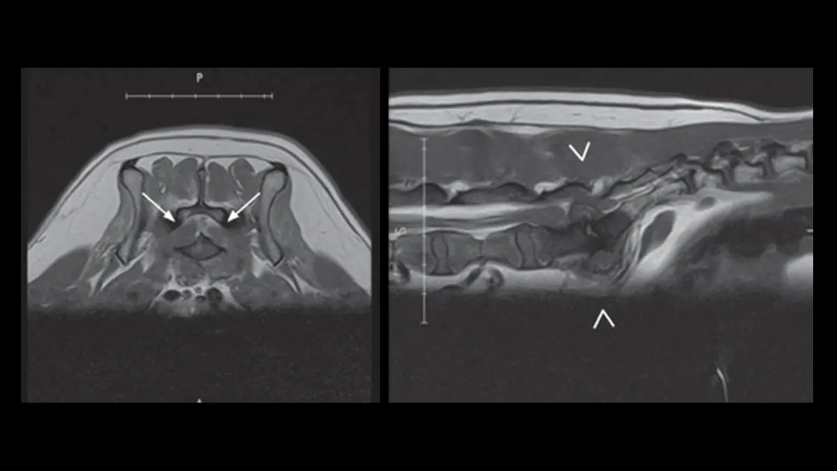
The picture at the top of this page shows two MRI slices from a 7-year-old German shorthaired pointer with weak back legs, a limp tail and a sore back. On the left, in cross-section, the two arrows point at swollen L7 nerve roots. On the right, in a side-on view, the arrowheads mark the L7-S1 level where a disc has pushed into the canal and presses on the cauda equina. This is a marked example, since swollen nerve roots are not found in every dog.
Radiologists look at:
- The L7-S1 disc: whether it has dried out (on T2 pictures a healthy disc looks bright in the middle and a worn one looks dark) and whether it bulges or has pushed into the canal
- The cushion of fat around the nerves: a pale ring normally surrounds the cauda equina, and where it has been squeezed out the nerves are being compressed
- The ligaments, joints and nerve exits: a thickened ligament or overgrown joints narrowing the tunnel, and whether an exit is pinched, which can explain a limp in just one back leg
- The alignment: whether L7 sits slightly out of line with the sacrum, which suggests instability
- The nerves themselves, which may look swollen or take up contrast dye (a dye injected into a vein that makes inflamed tissue stand out), and whether an infection or a tumor is behind the signs
Two honest caveats. Disc wear and mild bulging at this joint are very common in older large dogs with no signs, so your vet matches the scan to the examination instead of treating the picture alone. And a dog under anesthetic lies still and relaxed, which can make the squeeze look smaller than it is when the dog is up and moving.
Treatment: rest and medication, or surgery
There are two broad routes, and the choice depends on how much pain and nerve damage your dog has, what the scan shows, and your dog's age, job and general health.
Conservative (non-surgical) care suits dogs with pain and mild signs and no loss of bladder or bowel control. It means strictly limited activity for several weeks and then short lead walks, plus pain relief and anti-inflammatory drugs prescribed by your vet (often with a medicine aimed at nerve pain), weight loss if needed and physiotherapy. Many centers also offer an epidural steroid injection next to the nerves, which helps some dogs, though the benefit can wear off and signs can return once normal activity resumes.
Surgery aims to take pressure off the nerves. The usual operation is a dorsal laminectomy: the surgeon removes a window of bone over the tunnel, sometimes with part of the bulging disc or a thickened ligament, to give the nerves room, and may hold L7 and the sacrum together with screws and bone cement if the joint is unstable. It is major surgery, best done by a surgeon or neurologist who does it regularly. Most dogs improve in pain and in how they walk, though recovery takes weeks of strict rest. Risks include anesthetic and wound problems, and trouble can return or appear at a neighboring joint.
Many dogs treated before they lose bladder and bowel control do well, but dogs that have already lost control of urine or stool recover less predictably, so vets and neurologists advise against leaving severe signs untreated for long. Lumbosacral disease usually lasts for life, so some dogs need further treatment over time, and your vet or a neurologist can tell you what is realistic for your dog.
Looking after your dog at home
- Keep exercise calm and on a lead, with no jumping or rough play, use ramps and non-slip rugs, and keep your dog lean
- Use a harness instead of a collar, lift your dog with one arm under the chest and the other under the hind end, and never pull or lift by the tail
- Never give human painkillers such as ibuprofen or acetaminophen (paracetamol), which can be toxic to dogs, and give only what your vet prescribes
Why a second read can help
A lumbosacral MRI is a stack of detailed slices of a small, crowded area, and a lot rests on judgment: how much the canal is really narrowed, whether the nerve exits are pinched and whether a disc or a thickened ligament is the main culprit. Before you commit, you can ask a veterinary radiologist to re-read the MRI you already have.
DocOrbit offers a veterinary radiology second opinion online: a veterinary radiologist re-reads your dog's own MRI, and you can ask what the findings mean for treatment. A radiologist cannot examine your dog or feel how its back legs and tail respond, so this gives you and your vet another careful look at the same images and does not replace the vet or neurologist who treats your dog.
Is cauda equina syndrome in dogs an emergency?
Sometimes. A dog that is sore and stiff but still walking and passing urine normally can be seen within a day or two. It is an emergency if your dog suddenly cannot use its back legs, cannot pass urine or dribbles constantly, has new accidents with urine or stool, or is in severe pain, so call a vet right away if that happens.
Will my dog need surgery for lumbosacral disease?
Not always. Dogs with pain and mild signs can often be managed with rest, medication and sometimes an epidural injection, while surgery is usually advised for dogs with severe pain, weakness or loss of bladder or bowel control, or whose signs keep coming back. Your vet or a neurologist can say which route suits your dog.
Key takeaways
- Lumbosacral disease (cauda equina syndrome) is a narrowing at the joint between the last lower-back bone and the sacrum that squeezes the nerves to the tail, the bladder and bowel and the back legs
- It is most often seen in middle-aged and older large dogs, especially German Shepherds, as back pain, weak back legs and a low tail
- MRI shows the nerves and the cause of the squeeze, which an X-ray cannot, and is always matched to the examination
- Mild cases may be managed with rest and medication, severe or worsening cases often need surgery, and a dog that suddenly cannot use its back legs, cannot pass urine or has new accidents needs a vet today
Can I ask a veterinary radiologist about my dog's MRI report online?
Yes. Online veterinary radiology second opinions run entirely from home: you upload your dog's images, with no CD to post and no second trip to a specialty clinic, and a subspecialist veterinary radiologist re-reads the study and writes a structured report you can give to your own vet. DocOrbit returns that signed second read in 24 to 48 hours, and you can message the veterinary radiologist afterward with questions about the MRI and about what it means for your dog.
This article is for general information only and is not veterinary advice. Always discuss your animal's imaging results and next steps with a qualified veterinarian.