If your vet said "elbow dysplasia" after your young dog started limping on a front leg, or the X-ray report mentions a "fragmented coronoid process" or "osteoarthritis of the elbow," it is natural to worry. The reassuring part first: elbow dysplasia is one of the most common developmental joint problems in larger dogs, it is not an emergency, and although it usually cannot be cured, most dogs can be kept comfortable for years with the right plan. Here is what the X-ray shows, which signs to watch for and how the condition is managed.
What "elbow dysplasia" means
The elbow is where three bones meet: the upper arm bone (humerus) and the two forearm bones (radius and ulna). They must fit together like puzzle pieces for the joint to glide. "Dysplasia" means abnormal development: the bones grow unevenly, or a small piece of bone or cartilage fails to form properly, so the joint wears unevenly and arthritis follows.
It is an umbrella term, not a single disease. Your vet may name one or more of these:
- Fragmented medial coronoid process (FCP), the most common: a small bony hook on the inner side of the ulna cracks into pieces and rubs on the cartilage opposite it
- Osteochondritis dissecans (OCD): a flap of cartilage lifts off the rounded end of the upper arm bone, leaving a rough crater
- Ununited anconeal process (UAP): a small piece of the ulna at the back of the joint never fuses with the rest of the bone as the puppy grows, reported most often in German Shepherds
- Joint incongruity: the bones do not line up perfectly, leaving a small "step" so the load falls unevenly on the cartilage
Two or more can occur together, and in many dogs both elbows are affected.
Which dogs are affected, and why
It is a problem of medium, large and giant breeds. Labrador Retrievers, Golden Retrievers, Rottweilers, Bernese Mountain Dogs, German Shepherds, Newfoundlands, Saint Bernards and Chow Chows are reported most often, and it is uncommon in small breeds. The tendency is inherited, so responsible breeders have both parents' elbows X-rayed before breeding, and if you are choosing a puppy it is worth asking to see those scores. Fast growth may add to the risk, so keep a growing large-breed puppy lean. None of this means you did something wrong. The same breeds are also prone to hip dysplasia and a cruciate ligament tear, so a vet often looks at the whole dog rather than one joint.
Signs owners notice
Signs usually appear between about four and twelve months of age, as growth speeds up and the joint takes more load. Some dogs are only noticed as adults, when arthritis causes stiffness.
- A front-leg limp that is worse after exercise or after rest and often eases once the dog has warmed up
- Stiffness on getting up, and reluctance to play, jump or use stairs
- A short, stilted stride, with the elbows held out and the paws turned outward when standing
- A swollen or warm elbow, or pulling the leg away when it is bent
When both elbows are affected, which is common, there may be no obvious limp, because there is no healthy leg to compare. The dog just seems stiff, slow to get going and less keen than other dogs of the same age.
Elbow dysplasia is not an emergency, but a front-leg limp always needs examining, because other problems look similar: shoulder problems, growing-bone pain called panosteitis and fractures in young dogs, and occasionally a bone tumor in an older large dog, as our guide to osteosarcoma on a limb X-ray explains. See your vet today if your dog suddenly will not put the paw down, cries out when the leg is touched, has a hot, swollen elbow or has had a fall.
What elbow dysplasia looks like on an X-ray
The elbow is a tricky joint to X-ray: the bones overlap, and the coronoid process, the piece most often fragmented, hides behind the radius on a standard side view. Vets therefore take several views, almost always of both elbows even when only one leg is limping, including a flexed side view with the elbow bent. Dogs usually need sedation or a short anesthetic for good positioning.
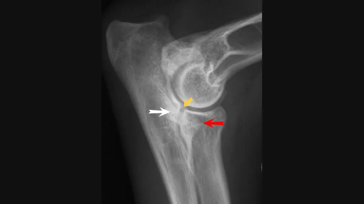
The picture at the top of this page is a flexed side view. The yellow arrow marks a step between the radius and ulna, the red arrow a missing front edge of the coronoid process, and the white arrow arthritic bone change on the ulna. Radiologists look for:
- Bone spurs (osteophytes) at the joint edges, especially on the anconeal process, radius and coronoid process, often the earliest and most reliable sign of strain
- Sclerosis, a band of denser, whiter bone just under the joint surface of the ulna
- A blunted, fuzzy or irregular coronoid process, or a fragment you can actually see
- A dark line separating the anconeal process from the ulna (UAP), or a flattened area or small defect on the inner side of the upper arm bone (OCD)
The honest catch is that an X-ray often shows the arthritis the problem has caused rather than the fragment itself, so a normal-looking X-ray does not completely rule out elbow dysplasia in a dog whose signs fit. A CT scan shows the coronoid process in cross-section and is more sensitive, and arthroscopy, a camera passed into the joint through a small incision, lets a surgeon see the cartilage and treat the problem in the same procedure.
How elbows are graded
Breeders use elbow scores to choose breeding dogs. The International Elbow Working Group (IEWG) scale runs from grade 0 (normal) to grade 3 (severe), and the Orthopedic Foundation for Animals (OFA) in North America uses normal, then grade I to III. Both mostly reflect how much arthritis has built up, so a young dog with a hidden coronoid problem can score as normal, and a grade is a breeding tool, not a prognosis.
Managing elbow dysplasia without surgery
The best plan depends on your dog's age, the specific problem and how sore the elbow is, and for many dogs the first step is not an operation.
- Weight control: a lean dog loads the elbows less, and it is the most effective thing you can do at home
- Controlled exercise: short, steady lead walks and swimming instead of ball chasing, repeated jumping and long forced runs
- Physiotherapy and hydrotherapy to keep muscles strong with little joint impact
- Joint supplements such as omega-3 fatty acids, which many vets recommend
- Pain relief prescribed by your vet, usually a veterinary anti-inflammatory. Never give human painkillers such as ibuprofen or paracetamol, which can be toxic to dogs.
When surgery is considered
Surgery comes into the picture when pain is not controlled by conservative care, or in young dogs with a clear fragment or flap irritating the joint. The options depend on the problem found.
- Arthroscopy: removing loose fragments and flaps and smoothing damaged cartilage through small incisions, mostly for FCP and OCD
- For UAP, fixing the loose piece with a screw or removing it
- Ulnar osteotomy: cutting the ulna so the bones align better in dogs with a step at the joint
- Larger rebuilds, such as humeral osteotomy or elbow replacement, at specialist centers for severely worn joints
Surgery can remove the source of irritation and ease pain, but it cannot reverse arthritis that has already formed, and most dogs still need some long-term management. Studies comparing surgery with careful non-surgical care have not shown one approach to be clearly better for every dog, so this is a decision to make with your vet or a surgeon.
Why a second read can help
Elbow X-rays are among the harder ones to read, and several calls are judgment calls: whether a fuzzy coronoid process is a fragment or just overlapping bone, whether a spur is mild or moderate, and whether a "normal" score hides a problem. Each can change the plan, which may involve CT, arthroscopy or an operation. Before you commit, you can ask a veterinary radiologist to re-read the X-rays you already have.
DocOrbit offers a veterinary radiology second opinion online: your dog's own X-rays are re-read by a veterinary radiologist, and you can ask what the findings mean for treatment and what to watch for. It gives you and your vet another careful look at the same images and does not replace the vet treating your dog.
Does elbow dysplasia always need surgery?
No. Many dogs are managed without an operation, using weight control, controlled exercise, physiotherapy and pain relief prescribed by a vet. Surgery is considered more often in young dogs with a clear fragment or cartilage flap, or when pain is not controlled by conservative care.
Can a normal X-ray rule out elbow dysplasia?
Not completely. A small coronoid fragment can be invisible on an X-ray, and the earliest changes in a young dog may be too subtle to see. If your dog's signs fit and the X-rays look normal, your vet may repeat them with more views, recommend a CT scan, or suggest arthroscopy.
Can a dog with elbow dysplasia live a normal life?
Many do. Elbow dysplasia is usually managed rather than cured, so the goal is comfort and good movement rather than a perfect X-ray. With a lean body weight, sensible exercise and treatment of pain as it appears, many affected dogs stay active for years.
Key takeaways
- Elbow dysplasia is an umbrella term for developmental elbow problems, such as a fragmented coronoid process, a cartilage flap (OCD) or joint incongruity, and it often affects both elbows
- It mostly affects medium and large breeds and shows as a front-leg limp or stiffness at four to twelve months; a sudden severe limp needs a vet the same day
- X-rays often show bone spurs rather than the fragment itself, so a normal-looking X-ray does not completely rule it out
- Management ranges from weight control and pain relief to surgery, and many dogs stay comfortable for years
Can I ask a veterinary radiologist about my dog's elbow X-rays online?
Yes. Online veterinary radiology second opinions run entirely from home: you upload your dog's images, with no CD to post and no second trip to a specialty clinic, and a subspecialist veterinary radiologist re-reads the study and writes a structured report you can give to your own vet. DocOrbit returns that signed second read in 24 to 48 hours, and you can message the veterinary radiologist afterward with questions about the X-rays and about what they mean for your dog.
This article is for general information only and is not veterinary advice. Always discuss your animal's imaging results and next steps with a qualified veterinarian.
