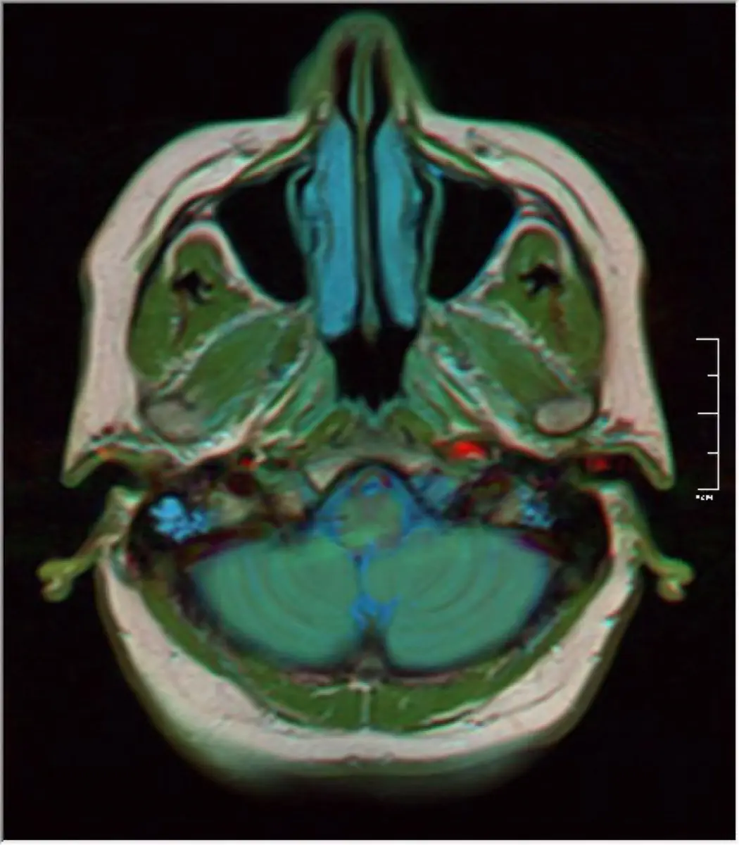
Watching your dog have a seizure is one of the most frightening moments an owner can face, and the very first fear that follows is often the word tumor. Take a breath: in most dogs, especially younger ones, a seizure does not mean a brain tumor. This article explains what actually causes seizures in dogs, when a brain MRI is the right next step, and what that scan can and cannot tell you.
What a seizure is, and why it happens
A seizure is a burst of abnormal electrical activity in the brain. In dogs it can look like full-body convulsions with paddling legs and loss of consciousness, or something subtler such as staring, facial twitching, or sudden collapse. Frightening as it looks, a single short seizure is rarely dangerous in itself. What matters is finding out why it happened.
Causes fall into three broad groups:
- Outside the brain — low blood sugar, liver or kidney problems, toxins, or electrolyte imbalances. Blood tests screen for these first.
- Inside the brain, structural — a tumor, inflammation (encephalitis), a stroke, or a malformation. This is what an MRI is designed to find.
- Idiopathic epilepsy — recurring seizures with a normal brain and normal blood work. This is the single most common cause in young and middle-aged dogs.
Does a seizure mean a brain tumor?
For most dogs, no. Age is the biggest clue. A dog that has its first seizure between roughly one and six years of age, with a normal neurological exam in between, most often has idiopathic epilepsy, a manageable lifelong condition rather than a tumor. The concern shifts when a seizure appears for the first time in an older dog, when seizures are paired with other signs such as circling, a head tilt, behavior changes, or vision loss, or when a dog does not fully return to normal between episodes. Those patterns raise the chance of a structural cause and are the reason a vet recommends imaging.
When your dog needs an MRI
An MRI is not automatic after a single seizure. Vets generally recommend it when:
- The first seizure happens under one year or over about six years of age
- Seizures come in clusters or are hard to control
- There are neurological signs between seizures
- Blood tests have not explained the cause
MRI is the best tool for looking at the brain's soft tissue, far better than X-ray or ultrasound, which cannot see through the skull in useful detail. It is the same reason MRI is the modality of choice for the canine spine; our explainer on intervertebral disc disease (IVDD) on MRI in dogs walks through how that works.
What the MRI can show
A brain MRI gives the veterinary neurologist a detailed map of the brain and can reveal:
- A mass and its location, size, and relationship to nearby structures
- Inflammation of the brain, which in dogs is a common and often treatable cause of seizures
- Evidence of a prior stroke or bleed
- Hydrocephalus (fluid buildup) or a structural malformation
- A completely normal brain, which supports a diagnosis of idiopathic epilepsy
The most common brain tumor in dogs is a meningioma, which grows from the membranes covering the brain and is often relatively slow-growing. The MRI helps the specialist estimate what a mass is likely to be, but the label is an educated read of the images; a definitive diagnosis usually needs a tissue sample. Assessing a suspicious mass is a theme across veterinary imaging, and our post on a splenic mass on ultrasound in dogs covers the same benign-versus-serious question in the abdomen.
Is it serious, and what happens next?
The honest answer depends on what the scan shows, and the range is wide. A normal MRI is genuinely good news and usually means epilepsy that can be controlled well with medication for years. Inflammation of the brain is serious but frequently responds to treatment. When a tumor is found, options are broader than many owners expect and depend on type and location: anti-seizure medication for comfort, corticosteroids to reduce swelling, and in selected cases surgery or radiation therapy. Many dogs with a slow-growing meningioma go on to have good quality time with the right plan. The point of the MRI is precisely to turn a terrifying unknown into a specific situation with specific choices.
Living with a dog that has seizures
Whatever the cause, a few practical habits help. Keep a simple seizure diary noting date, length, and what the episode looked like; this pattern is genuinely useful to your vet. During a seizure, keep your hands away from the mouth, move furniture out of the way, dim the room, and time it. A seizure lasting more than about five minutes, or repeated seizures without recovery in between, is an emergency and needs a vet right away. Give any prescribed medication consistently, as missed doses are a common reason seizures return.
Why a second read can help
Brain MRIs are detailed and open to interpretation, and distinguishing a tumor from inflammation, or judging whether a small change is meaningful, can shape a very different treatment plan. An independent expert read gives you confidence that the images have been interpreted carefully before you commit to surgery, radiation, or long-term medication. DocOrbit offers a veterinary second opinion on your animal's imaging that you can share with your own vet or neurologist, which can be reassuring when you are weighing surgery, radiation, or long-term medication for a beloved pet.
Does a seizure mean my dog has a brain tumor?
Usually not. In young and middle-aged dogs, the most common cause of recurring seizures is idiopathic epilepsy, where the brain looks structurally normal. A brain tumor becomes more likely when seizures start for the first time in an older dog or come with other neurological changes, which is exactly the situation where an MRI is most helpful.
When does a dog with seizures need an MRI?
An MRI is usually recommended when a dog has its first seizure under one year or over about six years of age, when seizures happen in clusters, when there are neurological signs between seizures, or when blood tests have not explained the cause. For a healthy young adult dog with a single typical seizure and normal exam, a vet may reasonably monitor first.
Are all brain tumors in dogs cancer?
No. The most common brain tumor in dogs is a meningioma, which arises from the membranes around the brain and is often slow-growing. Many meningiomas can be managed and, in the right cases, surgically removed or treated with radiation. The MRI helps estimate the tumor type, but a definitive diagnosis usually needs a tissue sample.
Does my dog need anesthesia for a brain MRI?
Yes. Dogs must stay completely still for a brain MRI, so it is performed under general anesthesia. Your vet will run pre-anesthetic checks first. Modern veterinary anesthesia is routine, and the imaging team monitors your dog closely throughout the scan.
What else can an MRI find besides a tumor?
An MRI can reveal inflammation of the brain (encephalitis), a prior stroke, a bleed, hydrocephalus, or a structural malformation, among other causes. It can also come back normal, which points toward idiopathic epilepsy. Ruling conditions in and out is a major reason the scan is worthwhile.
Key takeaways
- A seizure is frightening but, especially in younger dogs, most often points to idiopathic epilepsy rather than a tumor.
- Age and accompanying neurological signs decide whether an MRI is needed; older dogs with new seizures are checked more carefully.
- A brain MRI can find a tumor, inflammation, a stroke, or a normal brain, turning uncertainty into a clear plan.
- The most common canine brain tumor, meningioma, is often slow-growing and, in many cases, treatable.
Can I ask a veterinary radiologist about my pet's MRI report online?
Yes. Online veterinary radiology second opinions run entirely from home: you upload your images, with no CD to post and no second trip, and a subspecialist veterinary radiologist re-reads the study and writes a structured report you can give to your own vet. DocOrbit returns that signed second read in 24 to 48 hours. You can then message the veterinary radiologist with questions about the MRI and about what the report actually means for your pet.
This article is for general information only and is not veterinary advice. Always discuss your animal's imaging results and next steps with a qualified veterinarian.
