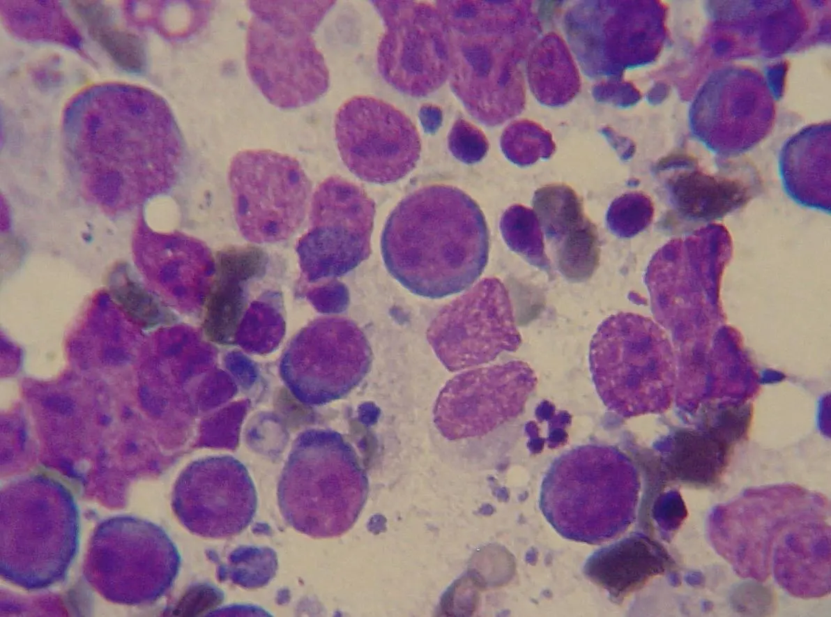
You were scratching your dog under the chin and felt something firm that should not be there, or your vet found it during a routine exam: a lump that turns out to be an enlarged lymph node, and then another, and another. The word that follows, lymphoma, is frightening, but an enlarged lymph node is not automatically cancer, and even when it is, dogs with lymphoma often respond well to treatment. Here is what a swollen node actually means, how vets confirm what it is, and why the whole body gets scanned once lymphoma is diagnosed.
What "enlarged lymph nodes" means
Lymph nodes are small, bean-shaped filters scattered through the body as part of the immune system. On a dog you can normally feel a few of them if you know where to press: under the jaw (mandibular), in front of the shoulders (prescapular), in the armpits (axillary), in the groin (inguinal), and behind the knees (popliteal). Healthy nodes are small and easy to miss. When several of these swell at once, painlessly, often to the size of a marble or a grape, that pattern is called multicentric lymphoma, and it is by far the most common form of the disease in dogs.
Less common forms involve the intestines (alimentary, with vomiting or diarrhea), the chest (mediastinal, which can affect breathing), the skin (cutaneous, with lumps or scaly patches), or a single organ (extranodal). Multicentric disease, the swollen-glands-under-the-jaw presentation, is what brings most owners in, and it tends to affect middle-aged to older dogs, though it can occur at any age. Some breeds show up more often than others, including Golden Retrievers, Boxers, Bullmastiffs, Basset Hounds, Scottish Terriers, and Rottweilers, but breed and age never replace an actual sample.
Reactive lymph nodes vs lymphoma: how vets tell the difference
Swollen lymph nodes are common, and most of the time they are not cancer. A node near an infected tooth, a healing wound, an ear infection, or even a recent vaccine can enlarge as the immune system does its job, and it usually settles once the underlying problem does. This is called reactive lymphadenopathy, and it is the everyday explanation for a swollen gland.
A few features shift the suspicion toward lymphoma rather than a reactive node:
- Several node regions enlarged at the same time, rather than just one near an obvious source of irritation
- The nodes feel firm and rubbery, and move freely under the skin rather than being tender or warm
- There is no obvious infection, wound, or dental problem nearby to explain it
- The swelling does not go down over a week or two
None of this is a diagnosis on its own. A reactive node and an early lymphoma node can genuinely look and feel alike, which is why the next step is a sample, not a guess.
Confirming the diagnosis
The usual first step is a fine needle aspirate: a thin needle draws a small sample of cells from the node, which are examined under a microscope. It is quick, rarely needs sedation, and often gives an answer the same day. A monotonous sheet of abnormal lymphocytes is highly suggestive of lymphoma; a mixed population of cells points back toward a reactive node or an infection instead.
When the aspirate suggests lymphoma, most oncologists recommend a biopsy, either a full node removed surgically or a larger needle-core sample, examined by a pathologist. This confirms the diagnosis and grades it, and it is usually paired with immunophenotyping, a test that sorts the cancer into B-cell or T-cell lymphoma. That distinction matters a great deal: roughly two-thirds of canine lymphomas are B-cell, which on average responds better and for longer to chemotherapy than T-cell disease.
Staging: what those extra scans actually check
This is the part that surprises a lot of owners. The diagnosis is already made by the time staging starts, so why does the dog also need chest X-rays, an abdominal ultrasound, and bloodwork? Because "lymphoma" describes the type of cancer, not how far it has spread, and how far it has spread shapes both the treatment plan and the honest conversation about outlook. Vets use a staging system that runs from a single affected node (stage I) through generalized node involvement (stage III, where most dogs are by the time they are diagnosed) to spread into the liver or spleen (stage IV) or the bone marrow (stage V). A dog is also marked substage a if it feels well, or substage b if it has symptoms such as poor appetite or lethargy, and that substage affects the outlook about as much as the numbered stage does.
Expect some combination of the following once lymphoma is confirmed:
- Bloodwork and urinalysis — checks organ function before chemotherapy and looks for a raised blood calcium level, which shows up in some T-cell lymphomas
- Chest X-rays — look for enlarged lymph nodes inside the chest and, in the less common mediastinal form, a mass around the heart and airway
- Abdominal ultrasound — checks whether the liver, spleen, or internal lymph nodes are involved
- Bone marrow aspirate — usually reserved for dogs with abnormal bloodwork, or to confirm stage V disease
Staging rarely changes whether treatment goes ahead. What it changes is how confidently your vet or oncologist can talk about the likely course, and whether anything unexpected, an unrelated mass or an enlarged organ, needs its own attention before chemotherapy starts.
Symptoms owners notice
Many dogs with multicentric lymphoma look and act completely normal at first. The nodes are painless, so a dog with several swollen glands under the jaw may still be eating, playing, and wagging its tail, which is part of why owners are often caught off guard. When symptoms do appear, they tend to include:
- Lethargy or a general drop in energy
- Reduced appetite or weight loss
- Increased thirst and urination, sometimes linked to a raised calcium level
- Vomiting or diarrhea, more typical of the alimentary form
- Coughing or labored breathing, more typical of the mediastinal form
A dog with none of these signs and simply enlarged, painless nodes is actually the more common picture, and it is a reasonably good sign at diagnosis, not a reason to assume the finding is being taken less seriously.
Treatment, honestly
For multicentric B-cell lymphoma, a combination chemotherapy protocol, most often built around cyclophosphamide, doxorubicin, vincristine, and prednisone (commonly called CHOP), is the standard of care and gives the best chance of remission, meaning the nodes return to normal size and the dog feels like itself again. Dogs generally tolerate these drugs far better than people do: doses are lower, severe reactions are uncommon, and most dogs stay bright and interested in life throughout treatment.
Chemotherapy is not the only option. A simpler course of prednisone alone controls symptoms and shrinks the nodes for a shorter period, and is a legitimate choice when full chemotherapy is not right for your family or your dog. If lymphoma comes back after a first remission, a rescue protocol using different drugs is often tried next. What treatment cannot promise, whatever the protocol, is a permanent cure; what it regularly delivers, especially for B-cell disease, is a meaningful stretch of good-quality time.
Why a second read can help
Staging carries real weight because the treatment conversation rests on it. A chest film or an abdominal ultrasound is easy to under-read when it is one of many studies moving through a busy clinic day, and a small internal node or a subtle liver change can shift a stage without being obvious at a glance. Asking a veterinary radiologist to double-check those images is a small step that can bring real clarity before you commit to a protocol. DocOrbit offers a veterinary second opinion on your dog's staging scans that you can bring to your own vet or oncologist, in the same spirit as a careful second look at a splenic mass on ultrasound or an aggressive bone lesion on X-ray. It does not replace your treating veterinarian; it gives you and your vet another informed set of eyes when the plan really matters. If a needle sample instead found a benign skin lump, our piece on lumps on dogs and the needle test covers that far more common outcome.
Are enlarged lymph nodes in a dog always lymphoma?
No. Lymph nodes commonly swell in response to infection, a nearby wound, dental disease, or even a recent vaccination, and that kind of reactive swelling usually settles on its own. What raises real suspicion for lymphoma is painless, firm enlargement of several nodes at once, in a dog that otherwise seems well. A needle sample is what actually tells the two apart.
How is lymphoma diagnosed in dogs?
The first step is usually a fine needle aspirate of an enlarged node, a quick in-clinic sample that often needs no sedation and can be read the same day. If it shows the abnormal lymphocyte population typical of lymphoma, a biopsy and immunophenotyping (working out whether it is B-cell or T-cell) confirm the diagnosis and its grade, which shapes both prognosis and the treatment plan.
What do staging scans check for in canine lymphoma?
Staging looks beyond the swollen node to see how far the disease has spread. Chest X-rays check the lungs and the internal lymph nodes in the chest, an abdominal ultrasound checks the liver, spleen, and internal lymph nodes, and bloodwork plus a bone marrow sample check the blood and marrow. Together they set the stage, which guides the treatment plan and gives a more realistic picture of the outlook.
Is lymphoma in dogs curable?
Usually not in the sense of a permanent cure, but it often responds very well to treatment. Most dogs with the common B-cell multicentric form go into remission on a combination chemotherapy protocol, meaning the nodes shrink back to normal and the dog feels well again, often for many months. T-cell lymphoma and dogs with systemic symptoms at diagnosis tend to have a shorter response, which is exactly why staging and a firm diagnosis come before any promises about the outlook.
Which dogs are most at risk of lymphoma?
Lymphoma can affect any dog, but it is seen more often in middle-aged to older dogs and in certain breeds, including Golden Retrievers, Boxers, Bullmastiffs, Basset Hounds, Scottish Terriers, and Rottweilers. Breed and age raise the index of suspicion when nodes are found enlarged, but the diagnosis itself always comes from a needle sample or biopsy, not from breed alone.
Key takeaways
- Enlarged lymph nodes are common and are usually reactive, not cancer; a needle sample is what actually tells the difference
- Multicentric lymphoma, painless swelling of several nodes at once, is the most common form in dogs
- A fine needle aspirate suggests the diagnosis; biopsy plus immunophenotyping (B-cell vs T-cell) confirms it
- Staging with chest X-rays, abdominal ultrasound, and bloodwork shows how far the disease has spread and shapes the treatment conversation
- Chemotherapy, most often a CHOP-based protocol, is the standard treatment and commonly brings dogs into remission for months, especially with B-cell disease
How do I have my dog's staging scans reviewed by another veterinary radiologist online?
Yes. Online veterinary radiology second opinions run entirely from home: you upload your images, with no CD to post and no second trip, and a subspecialist veterinary radiologist re-reads the study and writes a structured report you can give to your own vet. DocOrbit returns that signed second read in 24 to 48 hours. You can then message the veterinary radiologist with questions about the scans and about what the staging actually means for your dog.
This article is for general information only and is not veterinary advice. Always discuss your animal's imaging results and next steps with a qualified veterinarian.