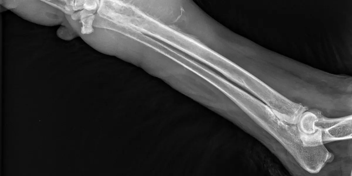
You took your dog in for a limp, expecting a sprain or the arthritis that creeps in with age. Instead you walked out with a word nobody is ready for: tumor. If the report says "aggressive bone lesion", "lysis", or "periosteal reaction", the shock is real, and so is the question you may not have said out loud: does this mean amputation? Here is what the radiologist is seeing, what else it could be, and what the choices look like.
What an aggressive bone lesion looks like on the X-ray
The word "aggressive" in a report is not a diagnosis. It describes behavior: the lesion has a poorly defined edge, it destroys bone faster than the body can wall it off, and the bone reacts in a disorganized way. Here is what is on the image:
- Lysis: bone being eaten away. It looks moth-eaten, or "permeative", as if riddled with tiny holes rather than cut with a clean edge
- New bone production: the tumor lays down disorganized bone, so destruction and building appear at once, a hallmark of osteosarcoma
- Cortical destruction: the hard outer shell of the bone is breached, not merely thinned
- Periosteal reaction: the bone lining throws down new bone in a hurry. Radiating spikes make the sunburst pattern, and the lifted lining can form a triangular shelf, the Codman's triangle
- Soft-tissue swelling: the tissues around the bone look thickened, matching the firm lump you may have felt
- The joint is usually spared: osteosarcoma tends to respect the joint space rather than cross into the neighboring bone. A lesion that crosses a joint points toward infection instead, a genuinely useful distinction
The classic locations and breeds
Osteosarcoma strongly prefers certain bones, and vets learn it with a phrase that sticks: away from the elbow, toward the knee. That means the distal radius (just above the wrist), the proximal humerus (near the shoulder), and the distal femur and proximal tibia (either side of the stifle, the dog's knee). It is largely a disease of big dogs: Great Danes, Rottweilers, Greyhounds, Irish Wolfhounds, and Dobermans are affected most often, and size matters more than breed alone. Most are middle-aged or older, though young large-breed dogs get it too. A classic location in a classic breed raises suspicion, but suspicion is not a diagnosis.
What else an aggressive lesion could be
This is the part most often skipped, and it matters most before anyone makes an irreversible decision. Osteosarcoma is the most common primary bone tumor in dogs, so it is a reasonable first thought. It is not the only one.
- Fungal osteomyelitis: some fungal infections invade bone and look almost identical to a tumor. Whether this belongs on the list depends on where you live and travel
- Bacterial osteomyelitis: bone infection, sometimes after an old wound, surgery, or a foreign body
- Bone cysts and other benign lesions: less common, but real, and not cancer
- Other primary bone tumors: chondrosarcoma, fibrosarcoma, and hemangiosarcoma of bone are treated differently
- Metastatic disease: cancer that started elsewhere and settled in bone
A radiologist weighs the pattern, location, breed, age, and clinical story, then gives a most-likely answer with a short differential list. What no radiologist can do is name the cell type from a radiograph.
The limp that would not go away
Most owners arrive because of a limp with a particular character: it does not resolve. Rest does not fix it. Anti-inflammatory medication may take the edge off and then stop working, which is worth telling your vet: a strain steadily improves, this does not. Other things owners notice:
- A firm, warm, painful swelling over one part of the leg, often at the wrist or shoulder
- Reluctance to jump into the car, climb stairs, or play
- Restlessness or pacing at night, sometimes the only sign of bone pain
- A sudden fracture after little or no trauma. A limb that breaks during ordinary activity is a red flag, because the bone was likely weakened from the inside
Persistent lameness in a large dog has many causes, and most are not cancer. Two common ones look similar from the outside: hip dysplasia on X-ray and a cruciate ligament tear. Both strike the same kind of dog that is also at risk of a bone tumor, which is why the X-ray is taken rather than assumed.
How the diagnosis is confirmed
The X-ray raises the question. Tissue answers it. Confirmation comes from a bone biopsy, or sometimes a needle sample (cytology, often taken with imaging guidance), examined under a microscope by a pathologist. That is what separates an aggressive tumor from a fungal infection, and one tumor type from another. Some teams proceed to surgery on strong imaging and clinical grounds when the picture is classic; many want histology first, especially when infection is plausible or when an owner needs certainty before consenting to an amputation. What is not defensible is treating the radiograph as if it were the pathology report.
Staging: why the chest matters more than the leg
This surprises almost every owner. Once a bone tumor is suspected, the most important images are of the chest, not the leg, because osteosarcoma tends to spread to the lungs, and whether the lungs are clear shapes the plan far more than the lesion in the limb. Expect some combination of:
- Chest radiographs, usually several views, to look for lung nodules
- Chest CT, because it picks up smaller nodules that plain films can miss
- Bloodwork, to check organ function and fitness for anesthesia
- Abdominal ultrasound or a bone scan in selected cases
Small lung nodules are easy to overlook, and they change everything. These are the most consequential images in the case.
Treatment, honestly
Amputation plus chemotherapy is the standard of care for confirmed limb osteosarcoma, and the reasoning is kinder than it first sounds. The affected bone hurts constantly, in a way medication only partly controls. Removing the limb removes that pain, and the relief is immediate. Chemotherapy afterwards targets microscopic disease that may already have reached the lungs.
The hardest part of this conversation is usually the owner's dread of the amputation, not the dog's experience of it. Veterinary surgeons say the same thing over and over: dogs cope with three legs far better than their owners expect. They do not mourn a limb; they notice that the pain has stopped, and most are up and walking within days. Very heavy or severely arthritic dogs need a closer assessment, and your surgeon will say so honestly.
Amputation is not the only path:
- Limb-sparing surgery: for selected dogs, most often when the tumor is in the distal radius
- Stereotactic radiation: a targeted technique that treats the bone while keeping the leg
- Palliative radiation and pain management: a humane, legitimate choice, not a failure. Good pain control is real treatment
What about the outlook? It depends: on whether the lungs are clear, where the tumor sits, and whether chemotherapy is used. A veterinary oncologist can give you realistic expectations once staging is complete.
Why a second read can help
Few situations in veterinary imaging carry this much weight, because the decision it drives is irreversible. A second expert read can separate an aggressive tumor from a fungal or bacterial bone infection, a different disease with a different treatment. It can catch subtle lung nodules on the staging chest films that change the whole plan. And it can confirm the lesion really is behaving aggressively before a leg is removed. DocOrbit offers a veterinary second opinion on your pet's imaging that you can bring straight to your own vet, surgeon, or oncologist. It does not replace your treating veterinarian; it gives both of you another informed set of eyes when it counts most. For a similar case, see our piece on a splenic mass on ultrasound.
Is every aggressive bone lesion on a dog X-ray osteosarcoma?
No. Osteosarcoma is the most common primary bone tumor in dogs, so it is a radiologist's first thought, but it is not the only thing that destroys bone. Fungal or bacterial osteomyelitis, bone cysts, other bone tumors such as chondrosarcoma, and cancer spread from elsewhere can all look aggressive. Tissue, not the X-ray, gives the final answer.
Can an X-ray alone diagnose osteosarcoma in a dog?
No. An X-ray shows that a lesion is behaving aggressively, where it sits, and how much bone has been lost, but it cannot identify the cell type. A biopsy or needle sample examined under a microscope is what confirms osteosarcoma and rules out the infections and other tumors that mimic it.
Does my dog definitely need an amputation?
Not automatically. Amputation with chemotherapy is the standard of care for confirmed limb osteosarcoma, mainly because the affected bone is genuinely painful and removing it ends that pain at once. Limb-sparing surgery and stereotactic radiation suit some dogs, and palliative radiation with good pain control is a legitimate choice. The decision is made with your veterinarian and an oncologist once the chest has been checked.
How do dogs cope after a leg amputation?
Far better than most owners expect, and veterinary surgeons say so consistently. Dogs do not grieve a lost leg the way people imagine; what they notice is that the constant pain is gone, and most are walking within days. Very heavy, very large, or severely arthritic dogs need a more careful conversation with your surgeon.
Why does my dog need chest X-rays if the tumor is in the leg?
Because osteosarcoma tends to spread to the lungs, and whether the lungs are clear shapes the plan far more than the leg does. Chest radiographs, or a CT scan that picks up smaller nodules, are part of standard staging before any irreversible surgery. Finding or excluding lung spread changes what your veterinarian recommends.
What are the early signs of a bone tumor in a dog?
The most common early sign is lameness that does not settle with rest or anti-inflammatory medication, usually in a large or giant breed dog. Owners often notice a firm, painful swelling over the leg, or restlessness at night. In some dogs the first sign is a sudden fracture through weakened bone after little or no trauma.
Key takeaways
- "Aggressive" on an X-ray describes behavior, not a diagnosis; only a biopsy or needle sample confirms what the lesion is
- Osteosarcoma is the most common primary bone tumor in dogs, but infection and other tumors can look identical
- Staging the chest matters more to the plan than the leg lesion
- Amputation with chemotherapy is standard, and dogs cope on three legs better than owners expect; limb-sparing surgery, radiation, and palliative care are real alternatives
- The decision is irreversible, which makes this a strong case for a second read
How do I have my pet's X-ray reviewed by another veterinary radiologist online?
Yes. Online veterinary radiology second opinions run entirely from home: you upload your images, with no CD to post and no second trip, and a subspecialist veterinary radiologist re-reads the study and writes a structured report you can give to your own vet. DocOrbit returns that signed second read in 24 to 48 hours. You can then message the veterinary radiologist with questions about the X-ray and about what the report actually means for your pet.
This article is for general information only and is not veterinary advice. Always discuss your animal's imaging results and next steps with a qualified veterinarian.