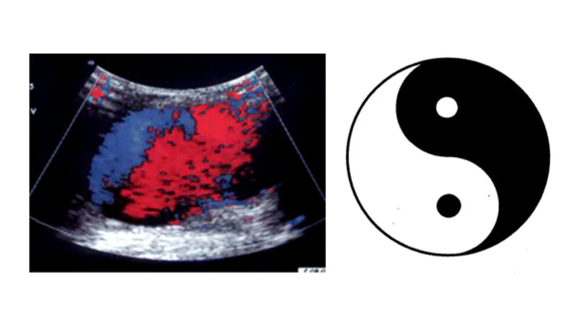
"Yin-yang sign" sounds like it wandered in from a philosophy class rather than a radiology report, but it is one of the more memorable named patterns in imaging. Seeing it on your ultrasound or CT report is not, on its own, alarming. It is a description of how blood is swirling in a small pocket, not a diagnosis. This article explains what radiologists mean by the term, where it typically shows up, what usually causes it, and what happens next.
What does "yin-yang sign" mean?
On color Doppler ultrasound, blood flowing toward the transducer is coded red and blood flowing away is coded blue. Most vessels carry a steady flow in one direction, so they stay mostly one color. When blood swirls back and forth within a confined pocket, flowing one way during part of the heartbeat and reversing during the other part, the red and blue regions curl around each other in a pattern that looks like the black-and-white halves of the yin-yang symbol.
The same idea turns up, less often, on CT or MR angiography, where swirling flow within a blood-filled sac creates two distinct zones of contrast enhancement that curve around each other instead of filling evenly. Some reports use "yin-yang sign" and "to-and-fro sign" interchangeably, since both describe the same underlying back-and-forth flow.
Where does the yin-yang sign usually show up?
The sign turns up in a fairly short list of situations, most of them centered on one problem: a small pocket of blood that connects to an artery through a narrow opening.
- A pseudoaneurysm, by far the most common cause. Blood leaks through a defect in an artery wall and is contained by surrounding tissue rather than by the vessel wall itself, forming a pulsating sac that fills and partially empties with each heartbeat
- A giant or partially thrombosed true aneurysm, where swirling flow persists in the part of the sac that is still open while the rest has clotted, a pattern also seen on scans looking for a brain aneurysm
- Occasionally, an arteriovenous fistula or another vascular connection with turbulent, bidirectional flow
Common causes
The finding itself is a description of flow, but the events that lead to it are fairly predictable:
- Arterial puncture for a cardiac catheterization or angiogram, most often at the groin (femoral artery) or wrist (radial artery) access site
- Blunt or penetrating trauma
- A liver, kidney, or pancreas biopsy that nicks a small artery
- Pancreatitis, when inflammation erodes into a nearby vessel
- Solid-organ transplant surgery or other recent abdominal or vascular surgery
- For a true aneurysm, the same wall-weakening processes that lead to aneurysms elsewhere: thinning of the vessel wall, high blood pressure, and, less often, an inherited connective tissue condition
Is it serious?
It depends on the size, location, and how fast it is growing, rather than on the label itself. A small pseudoaneurysm found by chance, especially at a puncture site after a recent procedure, is often stable and settles on its own with nothing more than monitoring. A larger or enlarging one, or one somewhere with less surrounding tissue to contain a leak, inside the abdomen for example, is treated more promptly because it can keep growing or rupture. As with any vascular finding, your own doctor weighs the sign together with your symptoms, the procedure or injury that came before it, and how it looks on follow-up imaging.
Symptoms
Many pseudoaneurysms cause no symptoms at all and are found incidentally on a follow-up scan or a Doppler ultrasound ordered for something else. When symptoms do occur, they can include:
- A pulsating lump under the skin, most often at a recent catheterization or biopsy site
- Localized pain, tenderness, or swelling
- A bruit, a soft whooshing sound a clinician can hear with a stethoscope over the area
- Rarely, sudden severe pain or a drop in blood pressure if the sac ruptures, which needs emergency care
How is it diagnosed and followed up?
Color Doppler ultrasound is the workhorse test, both for finding a pseudoaneurysm and for confirming that treatment worked, since it directly shows the swirling flow pattern. CT or MR angiography is used when the pseudoaneurysm sits somewhere ultrasound cannot reach well, such as deep inside the abdomen or chest, or when a treatment plan is being mapped out.
Management depends on size and behavior. Many small, superficial pseudoaneurysms, particularly right after a catheterization, close on their own and only need a repeat ultrasound to confirm the sac has clotted off. Larger, painful, or enlarging ones are usually treated directly, most often with an ultrasound-guided thrombin injection that clots the sac from the inside, or with compression of its neck under ultrasound guidance. Deeper or more complex pseudoaneurysms, and true aneurysms showing the same swirling pattern, may need catheter-based embolization or surgery. Follow-up imaging afterward confirms the flow pattern has resolved.
Why a second read can help
The yin-yang sign is easy to describe once you have seen it, but deciding how urgently it needs to be acted on takes real judgment: weighing the size of the sac, its location, how it is trending on prior scans, and the story behind it. An expert second read can help confirm the finding, put it into the context of your history, and give you something concrete to bring back to your own physician. DocOrbit offers an independent second opinion on your imaging that you can share directly with your care team. If you are weighing whether that is worthwhile, our guide on when to get a second radiological opinion covers how that decision usually gets made.
Does the yin-yang sign always mean I have a pseudoaneurysm?
Not always, but it is by far the most common explanation. The sign describes swirling, back-and-forth blood flow, and while a pseudoaneurysm is responsible in the large majority of cases, the same pattern can appear in a partially thrombosed true aneurysm or another turbulent-flow situation. Your doctor confirms which one it is by looking at where the finding sits and what led to the scan.
Is a pseudoaneurysm dangerous?
It depends mainly on its size and location. Small ones, especially at a recent puncture site, often resolve on their own with monitoring. Larger ones, or ones deeper in the body where a rupture would be harder to contain, are taken more seriously and usually treated directly rather than watched.
Does a yin-yang sign need treatment right away?
Not necessarily. Many small pseudoaneurysms close on their own within weeks and only need a follow-up ultrasound to confirm that. Ones that are large, growing, painful, or in a location where a rupture would be dangerous are typically treated sooner, often with a simple ultrasound-guided injection.
Can the yin-yang sign appear on CT or MRI, not just ultrasound?
Yes. It is most classically described on color Doppler ultrasound, but a similar swirling pattern of contrast can be seen on CT or MR angiography, particularly for pseudoaneurysms located deeper in the body or for aneurysms with a partially clotted, partially flowing sac.
What is the difference between a pseudoaneurysm and a true aneurysm?
A true aneurysm is a bulge in all three layers of a weakened artery wall, still contained by that wall. A pseudoaneurysm is not contained by the artery wall at all, it is a pocket of leaking blood held in only by surrounding tissue or clot, which is part of why it can behave differently and sometimes needs more prompt attention.
Key takeaways
- The yin-yang sign describes swirling, back-and-forth blood flow on color Doppler ultrasound, or a similar pattern on CT or MR angiography. It is a description of flow, not a diagnosis
- The most common cause by far is a pseudoaneurysm, often after a catheterization, biopsy, or injury
- Small, superficial pseudoaneurysms often resolve on their own; larger or deeper ones are usually treated directly
- Symptoms, when present, include a pulsating lump, localized pain, or a bruit heard with a stethoscope
- Follow-up ultrasound after treatment confirms the swirling flow pattern has resolved
Still unsure what the rest of your report says? Paste it into our free radiology report explainer and read it back in plain English, with each medical term defined and a short list of questions worth asking your doctor.
Can I get a second opinion on my ultrasound online?
Yes. Online radiology second opinions run entirely from home: you upload your images, with no CD to post and no second trip, and a subspecialist radiologist re-reads the study and writes a structured report you can give to your own doctor. DocOrbit returns that signed second read in 24 to 48 hours. You can then message the radiologist with questions about the ultrasound and about what the report actually means for you.
This article is for general information only and is not medical advice. Always discuss your imaging results and any next steps with a qualified physician.