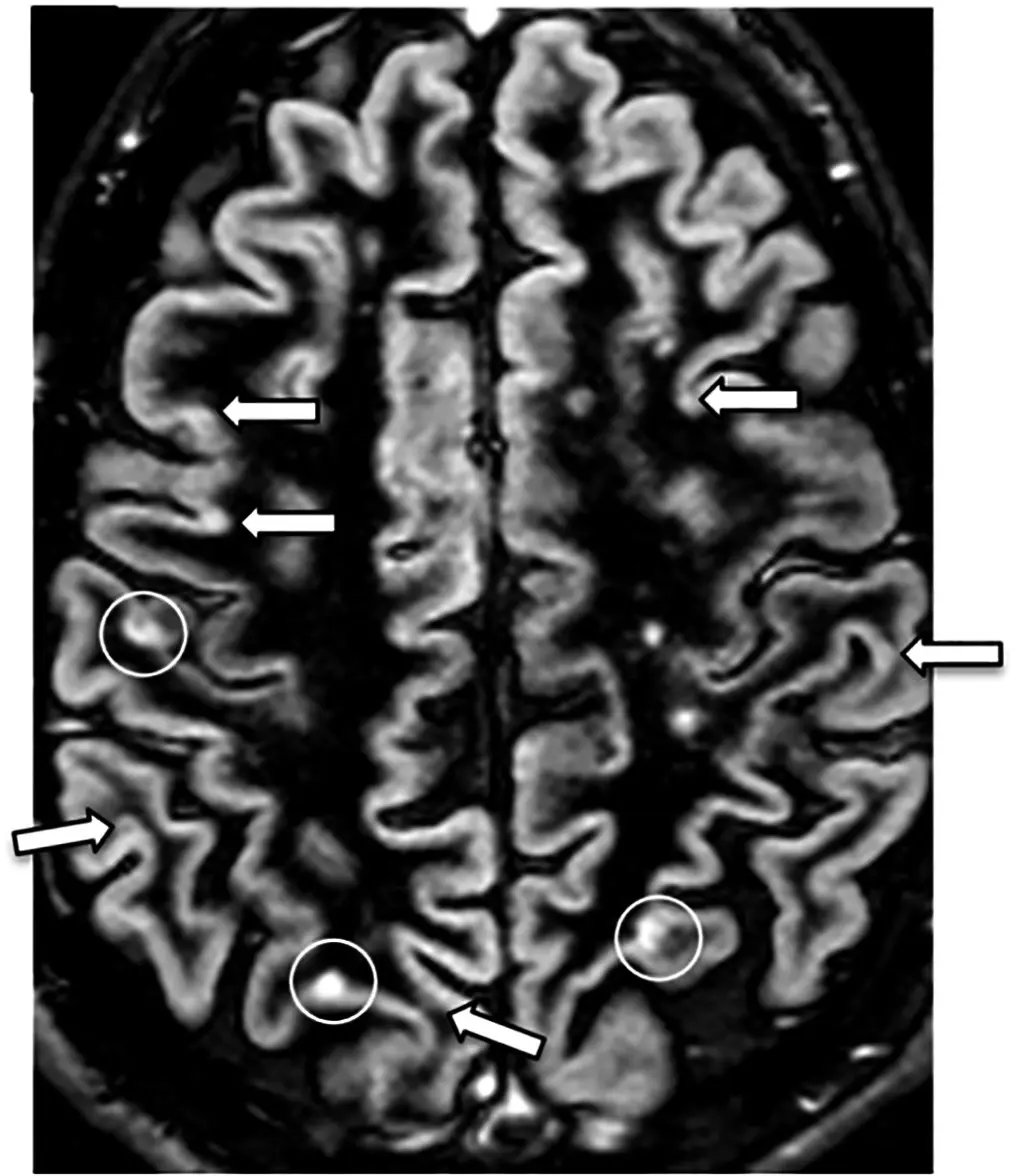
A lesion on an MRI report just means an area of tissue that looks different from the tissue around it, on its own it is not a diagnosis and it does not mean cancer. Radiologists use the word as a catch-all, from a harmless cyst to something that needs a closer look, and a good report describes what the lesion actually looks like rather than stopping at the word. Here is what radiologists mean by "lesion," what makes one worth a second look, and what typically happens next.
What radiologists actually mean by "lesion"
In radiology, a lesion is any area of tissue that appears abnormal compared with its surroundings, whether that is on an MRI, a CT, or an ultrasound. It is a description of appearance, not a cause. A lesion can turn out to be a fluid-filled cyst, a patch of scar tissue, an area of inflammation, a benign growth, or, less often, something malignant (cancerous).
Think of the word the way a proofreader uses a highlighter. Every highlighted word gets flagged for a second look, whether it turns out to be a typo, a stylistic choice, or nothing worth changing at all. "Lesion" works the same way in a report, it flags a spot that looks different from what surrounds it, without saying whether that difference matters.
What decides whether a lesion is worth worrying about
A radiologist weighs several features together, none of which settle it alone:
- Size, and whether it has grown compared with a prior scan
- Shape and borders, smooth and well-defined tends to reassure, irregular or blurred edges draw more attention
- How it behaves with contrast (a dye injected into a vein that makes blood vessels and some tissue light up, called enhancement)
- Where it sits, and whether that location is a common home for benign findings
- Your own symptoms and history, which images alone may not show
The same word can describe something trivial in one person and something needing a workup in another, the lesion is only part of the picture.
Common lesions that usually turn out to be nothing
Most lesions people ask about are, in the end, not dangerous. Simple cysts (fluid-filled pockets, common in the kidneys and liver) and hemangiomas (small, benign clusters of blood vessels, common in the liver) are picked up so often that radiologists barely comment on them beyond noting they look typical. Small scars, old healed injuries, and minor fatty deposits also get called lesions and need no action. A lung nodule found on a CT scan and a kidney cyst found on ultrasound walk through that same reasoning in more detail.
What happens after a lesion is found
What comes next depends on what the lesion looks like and where it is. Some lesions look confidently benign on imaging alone, and the report simply notes an incidental finding with no follow-up needed. Others are called indeterminate, meaning the appearance alone can't settle it, and a repeat scan in a few months is recommended, since staying unchanged is itself reassuring. A smaller number are referred for a biopsy (removing a small tissue sample to examine under a microscope) or a specialist visit. If the wording feels vague, sharing the images for a subspecialist second read, the kind DocOrbit provides, can put a clearer name on what your report calls a lesion.
Does a lesion mean I have cancer?
No, in the large majority of cases a lesion is not cancer. The word only describes an area that looks different from the surrounding tissue, and most turn out to be cysts, benign growths, scar tissue, or normal variation. Whether cancer is even on the list of possibilities depends on the lesion's appearance, its location, and your own history.
What's the difference between a lesion and a tumor?
A tumor is one specific type of lesion, an abnormal growth of tissue that can be benign or malignant. Lesion is the broader term and also covers things that are not growths at all, such as cysts, scars, and inflammation. Every tumor is a lesion, but most lesions are not tumors.
Why didn't the radiologist just tell me what it is?
Some lesions have a signature appearance a radiologist can name with confidence from the images alone. Others look similar to several different things, and telling them apart needs more information, such as a comparison with an older scan, blood work, or a biopsy. Calling something a lesion while that information is gathered is accurate, not evasive.
How soon is a lesion usually followed up with another scan?
There is no single timeline, intervals vary widely by the lesion's type, size, and location, and your own doctor sets the actual schedule based on your history. A follow-up in a few months is common for indeterminate findings, and some lesions are simply reassessed at the next scan you happen to need anyway.
Key takeaways
- "Lesion" describes appearance, not a cause, it is not a diagnosis by itself
- Most lesions found on MRI are benign, cysts and hemangiomas among the most common
- Size, growth, shape, and contrast enhancement all factor into how concerning one looks
- Follow-up ranges from none needed to a repeat scan to a biopsy, depending on the specifics
Can I ask a radiologist about my MRI report online?
Yes. Online radiology second opinions run entirely from home: you upload your images, with no CD to post and no second trip, and a subspecialist radiologist re-reads the study and writes a structured report you can give to your own doctor. DocOrbit returns that signed second read in 24 to 48 hours. You can then message the radiologist with questions about the MRI and about what the report actually means for you.
This article is for general information only and is not medical advice. Always discuss your imaging results and any next steps with a qualified physician.
