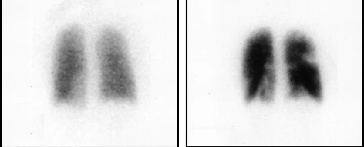
Somebody mentioned a possible clot in your lungs, and instead of the CT scan you were expecting, you were sent to nuclear medicine for something called a V/Q scan. You breathed in a gas or a fine mist, you had an injection, you lay under a large camera, and now a report has come back talking about mismatched defects and probability. Here is the honest starting point: this test is chosen precisely because it can answer the clot question safely in people for whom a CT is a poor fit, and most V/Q scans come back reassuring.
What a V/Q scan actually measures
V stands for ventilation, which is air. Q is the medical shorthand for perfusion, which is blood flow. The scan is really two short studies compared side by side.
In the ventilation part, you breathe in a small amount of a radioactive gas or an aerosol mist through a mouthpiece or mask. It travels wherever air travels, so the camera sees which parts of your lungs are being ventilated.
In the perfusion part, a tracer is injected into a vein in your arm. It follows the blood into the smallest lung vessels and lodges there briefly, so the camera sees which parts of your lungs are receiving blood.
Then the two maps are laid over each other. The whole diagnostic power of the test comes from one simple comparison: does air and blood reach the same places, or not?
Why you were sent here instead of for a CT
For most adults with a suspected pulmonary embolism, the first-line test is a CT pulmonary angiogram, which uses iodinated contrast dye to light up the lung arteries. A V/Q scan is chosen when that dye or that dose is best avoided. The common situations are:
- Pregnancy. A V/Q scan delivers a lower radiation dose to the chest and breast tissue than a CT pulmonary angiogram, which matters for the mother, and many departments modify the protocol further in pregnancy.
- Reduced kidney function. Iodinated contrast is filtered by the kidneys, so it is used cautiously when kidney function is already impaired.
- A previous reaction to contrast dye. No iodinated contrast is used in a V/Q scan at all.
- Younger patients, particularly younger women. Avoiding breast radiation over a lifetime of imaging is a legitimate consideration.
- A completely clear chest X-ray. V/Q scans are most reliable in lungs that are otherwise normal, which is often the case in exactly these patients.
None of these mean your case is being taken less seriously. Suspected pulmonary embolism is handled urgently regardless of which scan is used, and the choice of test is about matching the tool to the patient. If the concern started with a swollen leg, our explainer on deep vein thrombosis covers the other half of the same problem, since clots in the lungs usually start in a leg vein.
Matched, mismatched, and what a defect means
The word defect sounds alarming, but in this report it simply means an area where the tracer is not showing up as expected. It is a description of the image, not a verdict about your lungs. What matters is whether the two maps agree.
- Mismatched defect. Air gets in, blood does not. The airways in that segment are fine, but something is blocking its artery. This is the classic pattern of a pulmonary embolism, and it is the finding the test was designed to catch.
- Matched defect. Neither air nor blood reaches that area. Lung tissue that is not ventilated automatically has its blood flow reduced, so a matched defect usually points to a lung problem rather than a clot: pneumonia, collapse, emphysema, or fluid around the lung. Our article on pleural effusion covers one of the more common causes of a matched pattern at the lung base.
- Reverse mismatch. Blood flow looks better preserved than ventilation. This also generally points away from a clot and toward an airway or airspace problem.
Size and shape matter too. Radiologists look for defects that follow the natural wedge shape of a lung segment, because that is how arteries branch. A small, vague, non-segmental patch carries much less weight than a crisp wedge.
Low, intermediate, and high probability: why the report hedges
Very few tests in medicine report a probability rather than a yes or no, and seeing that word about your own lungs is unsettling. There is a reason for it. The original large study of this test, PIOPED, and its later refinement, modified PIOPED II, showed that certain patterns predict a clot far more reliably than others, but that no pattern is perfect. Rather than pretend to a certainty the images do not carry, radiologists report the strength of the evidence.
In broad terms:
- Normal. Perfusion is complete throughout. A truly normal perfusion scan is very good news and is generally accepted as ruling out a clinically significant pulmonary embolism.
- Low probability. The findings are much more likely to be explained by something other than a clot. Combined with a low clinical risk score, this usually ends the investigation.
- Intermediate or indeterminate probability. The pattern is genuinely ambiguous. This is more common in people who already have lung disease, and it means the scan cannot settle the question on its own.
- High probability. Two or more large mismatched segmental defects, or their equivalent. In a patient whose symptoms fit, this is treated as a pulmonary embolism.
You may also see PISAPED mentioned. That is a simplified approach in which the perfusion scan is read together with the chest X-ray and reported as either compatible or not compatible with pulmonary embolism, avoiding the middle category altogether. Some departments prefer it precisely because patients and referring doctors found the intermediate bucket so frustrating.
The key thing to understand is that no version of this report is meant to stand alone. It is deliberately designed to be combined with your clinical risk score, your D-dimer blood test, and any leg ultrasound. A low probability scan in a high-risk patient does not close the case, and an intermediate scan in a low-risk patient rarely leads anywhere alarming.
The tracer, the radiation, and pregnancy
The perfusion injection is usually technetium-99m bound to tiny albumin particles, which are protein particles that briefly lodge in the small lung vessels before being broken down and cleared. The ventilation part uses either technetium-99m as a fine aerosol or an inert radioactive gas. Both have short half-lives and leave the body quickly.
The total radiation dose from a V/Q scan sits in the same broad range as many routine imaging tests and is lower to the chest than a CT pulmonary angiogram. In pregnancy, the dose reaching the baby is very small, and it can be reduced further by lowering the injected activity, skipping the ventilation phase when the chest X-ray is clear, and encouraging fluids and frequent bladder emptying afterwards so the tracer clears faster.
It is entirely reasonable to feel uneasy about any radiation while pregnant. The framing that helps most patients is comparative: the risk from this scan is very small and theoretical, while an untreated clot in the lungs is a real and immediate danger to both mother and baby. That asymmetry is why the test gets done. For the wider picture, our guide to radiation dose in radiology scans puts the numbers in context.
What happens next, and the waiting
When a V/Q scan is ordered for suspected pulmonary embolism, it is an urgent study. Images are typically read the same day, often within a couple of hours, and in many cases treatment with a blood thinner has already been started before the result arrives, because doctors do not wait to protect a patient they consider at risk.
If the scan is normal or low probability and your risk score is low, the investigation usually stops there and attention turns to other explanations for your symptoms. If it is high probability, anticoagulation is confirmed and continued, typically for at least three months, with the exact plan depending on why the clot formed. If it comes back intermediate, expect one more step rather than bad news: a leg ultrasound, a repeat of the blood test, or a CT after all if it is now considered safe.
The waiting is often the hardest part, and it helps to know that the anxious period is usually measured in hours, not days, and that people are acting on your case in the background while you sit with the uncertainty.
Why a second read can help
V/Q scans are among the more interpretation-dependent studies in imaging. The same set of images can be called intermediate by one reader and low probability by another, and that difference can decide whether someone spends months on anticoagulation. It is a genuinely useful place for an independent look, particularly when the report is equivocal or when the result does not match how you actually feel. DocOrbit offers a subspecialty-matched second read you can take back to your own doctor, which patients most often find worthwhile when a borderline report is driving a long-term treatment decision. Our guide on when to get a second radiological opinion sets out the situations where it changes management.
Is a V/Q scan safe during pregnancy?
It is one of the tests specifically chosen for pregnancy, which tells you a lot. The radiation reaching the baby from a V/Q scan is very small, and in many centres the ventilation part is shortened or skipped in pregnant patients to lower it further. Doctors weigh that small exposure against the real risk of leaving a clot in the lungs undiagnosed, and an untreated clot is by far the bigger danger to both mother and baby.
What does a mismatched defect on a V/Q scan mean?
It means an area of lung is being ventilated normally with air but is not receiving its normal blood supply. That combination is the classic signature of a blood clot blocking a pulmonary artery branch, because a clot stops blood flow without affecting the airways. One or more mismatched defects of a certain size and shape is what pushes a report toward high probability of pulmonary embolism.
Why did I get a V/Q scan instead of a CT scan for a possible clot?
The usual reasons are pregnancy, reduced kidney function, a previous reaction to iodinated contrast, or a young patient in whom the breast and chest radiation dose from CT is worth avoiding. A V/Q scan answers the same clinical question without iodinated contrast and with a lower dose to the chest. It is a deliberate choice, not a downgrade or a sign that your case was treated as less urgent.
What does intermediate probability mean on my lung scan report?
It means the pattern is neither clearly normal nor clearly typical of a clot, so the scan alone cannot settle the question. This is a known limitation of the test rather than an error, and it happens more often in people with existing lung disease. Your team combines it with your symptoms, D-dimer, leg ultrasound findings, and clinical risk score, and may order a further test to reach a decision.
How long does it take to get V/Q scan results?
When the scan is done because a clot is suspected, it is treated as urgent and the images are usually read the same day, often within hours. If you are an inpatient or in the emergency department, the result frequently reaches your treating doctor before you are back in your bed. Outpatient scans arranged to investigate long-standing breathlessness are less time-critical and may take a few days.
Key takeaways
- A V/Q scan compares two maps of your lungs, one for air and one for blood flow, and looks for places where they disagree.
- A mismatched defect, meaning air present but blood absent, is the pattern that suggests a clot. A matched defect usually points to a lung problem instead.
- The probability wording reflects the honest limits of the test, not doubt about your care. It is designed to be read alongside your symptoms and blood tests.
- You were sent for this rather than a CT because of pregnancy, kidney function, contrast allergy, or dose considerations, not because your case was ranked as less urgent.
- The radiation dose is small, lower to the chest than a CT pulmonary angiogram, and an untreated clot carries far more risk than the scan does.
If the rest of your report reads like another language, our free radiology report explainer rewrites it in everyday words, defines the terms it uses and suggests what to ask at your next appointment.
Can I ask a radiologist about my nuclear medicine scan report online?
Yes. Online radiology second opinions run entirely from home: you upload your images, with no CD to post and no second trip, and a subspecialist radiologist re-reads the study and writes a structured report you can give to your own doctor. DocOrbit returns that signed second read in 24 to 48 hours. You can then message the radiologist with questions about the nuclear medicine scan and about what the report actually means for you.
This article is for general information only and is not medical advice. Always discuss your imaging results and any next steps with a qualified physician.
