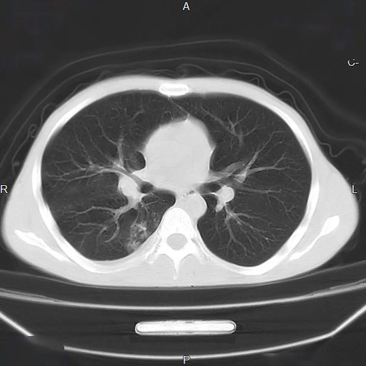
Finding the phrase "tree-in-bud pattern" on a chest CT report is disorienting, mostly because it sounds like it belongs in a gardening book rather than a medical document. Here is the reassuring starting point: tree-in-bud is a description of a shape the radiologist can see in the smallest airways, not a diagnosis, and in the large majority of people it points to an infection or inflammation that settles down. This article explains what radiologists mean by the term, what causes it, when it carries weight, and what usually happens next.
What does "tree-in-bud" mean?
Deep in the lung, the airways keep dividing into finer and finer branches until they reach tubes narrower than a human hair. Normally these tiny airways are invisible on a CT scan because they are filled with air and their walls are too thin to see. When something fills them, mucus, pus, fluid, or inflammatory cells, they suddenly become visible.
What appears is a small branching line with tiny round dots at the ends of the branches. To radiologists that shape looks like a budding twig in spring, which is where the name comes from. It is a thin-section CT finding, essentially never something you would see on a plain chest X-ray, because the structures involved are only a few millimetres across.
Several details about the pattern carry meaning, and a good report usually mentions them:
- Whether it is focal (one region) or spread through both lungs
- Which part of the lung it favours, since some causes have characteristic locations
- Whether it sits alongside other findings such as thickened airway walls, widened bronchi, or patches of denser lung
- Whether it has appeared, moved, or faded since any earlier scans
These details usually matter more than the phrase itself, because they are what point toward a cause.
Common causes
Tree-in-bud is the lung's way of showing that the small airways have something in them. Infection is by far the most common reason, and the pattern is often a sign that the body is actively dealing with something rather than that something is permanently wrong. Common causes include:
- Ordinary bacterial chest infections, including bronchopneumonia
- Viral chest infections and their aftermath, which can leave the pattern behind for weeks
- Mycobacterial infections, including tuberculosis and the slower-growing non-tuberculous mycobacteria
- Aspiration, when small amounts of food, drink, or stomach contents reach the airways, often during sleep or after reflux
- Bronchiectasis, where widened airways collect mucus that is hard to clear
- Long-standing conditions that impair mucus clearance, such as cystic fibrosis and primary ciliary dyskinesia
- Inflammatory airway disease linked to allergy, certain fungi, or a connective tissue condition
- Uncommonly, tumour cells travelling in the tiniest lung arteries, which produces a similar branching look
The same shape can therefore reflect a chest infection you are already recovering from or, much less often, something that needs a longer look. Context does the heavy lifting here. If your report also mentions hazy areas of lung, our explainer on what ground-glass opacity means covers a finding that often appears alongside it.
Is it serious?
For most people it is not the alarming finding it can sound like. A limited patch of tree-in-bud in someone who has recently had a cough and a fever is usually a straightforward, self-limited thing, and it commonly disappears on a follow-up scan without any specific treatment aimed at the pattern itself.
Doctors take a closer look when the pattern is widespread across both lungs, when it keeps coming back in the same area, or when it comes with weight loss, night sweats, coughing up blood, or a cough that has lasted months. Those combinations raise the possibility of a chronic infection such as tuberculosis or a non-tuberculous mycobacterium, which are treatable but need to be identified properly. As always, interpretation depends on your symptoms, exam, and history rather than the scan alone.
Symptoms
The pattern itself causes nothing. Any symptoms come from whatever is filling the airways. Depending on the cause, that might mean:
- A cough, dry or productive, which is the most common accompaniment
- Fever, chills, or simply feeling unwell during an active infection
- Shortness of breath or wheeze, especially if the airways are already narrowed
- A long-running cough with daily mucus, when bronchiectasis or a chronic infection is behind it
- No symptoms at all, when the finding turns up by chance on a scan ordered for something unrelated
The last case is more common than people expect. Small areas of tree-in-bud are regularly spotted on CT scans done for entirely different reasons, in people who feel completely fine.
How is it diagnosed and followed up?
Because the pattern is a clue rather than an answer, the next step is usually modest. When a recent or ongoing chest infection explains it, the most useful move is often to treat the infection and repeat the CT after several weeks, once the lung has had time to clear. A pattern that has resolved confirms it was temporary and closes the question.
When the picture is less straightforward, doctors may add sputum samples, including specific tests for tuberculosis and other mycobacteria, blood tests, or breathing tests. A referral to a respiratory specialist is common if the pattern is extensive, recurrent, or accompanied by a chronic productive cough. Bronchoscopy is occasionally used when a sample is needed and sputum has not given an answer, but it is far from routine. The same patient, unhurried approach applies to other chest findings, our guide to fluid around the lung shows how a finding gets read in context rather than acted on in a rush.
Why a second read can help
Tree-in-bud is one of those findings where the distribution and the company it keeps matter more than the phrase, and where a careful reader can meaningfully narrow the list of likely causes. A clear, expert read can help separate the resolving chest infection from the pattern that genuinely deserves sputum testing and a specialist opinion, and it gives you something concrete to bring to your own doctor. DocOrbit offers an expert second read of your scan that you can share directly with your care team, so the finding is understood in the context of your full history. If you are weighing whether to seek one, our guide on when to get a second radiological opinion may help, and you can read more broadly about the role of second opinions in radiology.
Does a tree-in-bud pattern mean I have tuberculosis?
Not on its own. Tuberculosis is one recognised cause of the tree-in-bud pattern, which is why radiologists mention it, but ordinary bacterial and viral chest infections produce the same appearance far more often. Doctors decide how likely tuberculosis is from your symptoms, your exposure history, where in the lung the pattern sits, and sputum testing, not from the CT picture alone.
Is tree-in-bud a sign of lung cancer?
Very rarely. Tree-in-bud reflects material inside the small airways, which is the signature of infection and inflammation rather than of a tumour. There is an uncommon version caused by tumour cells inside tiny lung arteries, but it is unusual and is normally considered only in someone already known to have cancer. For most people the pattern points somewhere far more ordinary.
Can a tree-in-bud pattern go away?
Yes, and it usually does. When the cause is an infection or a short-lived inflammation, the small airways clear as you recover and the pattern fades over weeks to a few months. That is exactly why a repeat CT after treatment is such a common next step, since a pattern that has disappeared needs nothing further.
How serious is a tree-in-bud pattern on a CT scan?
It depends on the cause, how much of the lung is involved, and whether it persists. A small patch in someone recovering from a chest infection is usually minor and temporary, while a widespread pattern, or one that stays put over months, is looked into more carefully. The term describes an appearance, not a severity, so it needs to be read together with your symptoms and history.
What is the follow-up for a tree-in-bud pattern?
Follow-up varies by patient and by suspected cause. When an ordinary infection is likely, treatment and a repeat scan after several weeks is often all that is needed. When the pattern is widespread, recurrent, or paired with a chronic cough, doctors may add sputum tests, breathing tests, or a referral to a lung specialist before deciding on any longer-term plan.
Key takeaways
- Tree-in-bud describes tiny branching lines with dots at their tips on a CT scan, caused by material filling the smallest airways. It is a description, not a diagnosis
- Infection and inflammation account for the large majority of cases, and cancer is a very rare explanation
- The pattern often clears on its own as the underlying infection resolves, which is why a repeat scan is a common next step
- Widespread or persistent patterns, especially with a long-running cough, night sweats, or weight loss, get a closer look including sputum testing
- Where the pattern sits and what else is on the scan matter more than the phrase itself, so interpretation always depends on your symptoms and history
Your report probably contains more than this one finding. Our free radiology report explainer puts the whole thing into plain English and gives you a short list of questions to take to your doctor.
Can I ask a radiologist about my CT report online?
Yes. Online radiology second opinions run entirely from home: you upload your images, with no CD to post and no second trip, and a subspecialist radiologist re-reads the study and writes a structured report you can give to your own doctor. DocOrbit returns that signed second read in 24 to 48 hours. You can then message the radiologist with questions about the CT and about what the report actually means for you.
This article is for general information only and is not medical advice. Always discuss your imaging results and any next steps with a qualified physician.