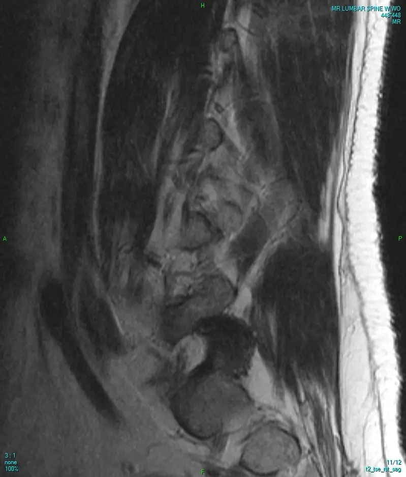
You already have the scan, a lumbar MRI for leg pain, a cervical MRI for hand numbness, or a thoracic MRI ordered along the way. A spine MRI second opinion is exactly what it sounds like: a radiologist who reads spine imaging every day reviews your existing images independently and sends back a written report you can bring straight to your own doctor. Maybe the report came back reassuring and something still does not sit right, maybe it flagged a finding you do not fully understand, or maybe you simply want to have your spine MRI re-read by someone who reads nothing else before you act on what it says. Here is what that second read covers, why the spine rewards the extra attention, and what happens after you send your images in.
A spine MRI holds more than the question it was ordered to answer
A single spine MRI, whether it covers the lumbar, cervical, or thoracic segment, produces well over a hundred images across several sequences, T1, T2, STIR, sometimes fat-saturated or contrast-enhanced series, each one showing the same vertebrae and soft tissue differently. In a busy radiology department, a general radiologist works through that stack alongside the day's chest X-rays, abdominal CTs, and everything else on the list, under real time pressure. The read is optimized to answer the specific clinical question on the request form, rule out a fracture, evaluate sciatica, follow up a known disc problem, not to comb every level of the spine for everything that could conceivably be there.
Small or incidental findings, a subtle annular tear at a level that was not the focus, an early Modic marrow change, a mildly narrowed foramen one level above the symptomatic one, are exactly what a fast first read is not looking for. None of this reflects any individual radiologist's competence; it is what volume and time do to any read. A slower, dedicated second read buys back the attention the first pass did not have room for.
Why a spine MRI benefits from both a neuroradiologist and an MSK radiologist
A general radiologist reads everything, chest, abdomen, bone, brain, spine, often in the same shift. Spine imaging is unusual in that it does not belong to one subspecialty. A neuroradiologist is trained to read the spinal cord itself and the nerve roots branching off it, catching a cord signal change or a subtle case of nerve root compression that a broader read might attribute to something else. A musculoskeletal (MSK) radiologist is trained to read the discs, facet joints, and bone marrow with the same depth, catching an early facet arthropathy or a marrow pattern that deserves a closer look. A proper spine MRI second opinion draws on both, rather than treating the spine as belonging to just one of them.
This is the logic behind a second opinion on a lumbar, cervical, or thoracic MRI from a subspecialist rather than a general reader: not carelessness on the first read, but a narrower, deeper field of view that statistically catches what a broad one is more likely to under-call.
What a spine MRI actually shows, structure by structure
A thorough spine MRI review works through the same anatomy a spine subspecialist is trained to check at every level, structure by structure:
- Vertebral bodies and bone marrow — checked for compression fractures, marrow edema patterns known as Modic changes, and incidental lesions such as a hemangioma, one of the most common findings in the spine
- The intervertebral discs — assessed level by level for degenerative disc disease and disc herniation, and for whether that herniation is displacing or touching a nerve root
- The facet joints — the small paired joints at the back of each vertebra, reviewed for facet arthropathy and for synovial cysts that can form alongside it
- The spinal canal — measured for central canal stenosis, narrowing that can come from disc bulging, facet overgrowth, and a thickened ligamentum flavum together
- The neural foramina and nerve roots — the small openings each nerve root exits through, checked for narrowing that can pinch a root and explain pain radiating down an arm or leg
- The spinal cord, on cervical and thoracic studies — reviewed for cord signal change, compression, a syrinx, or demyelinating plaques like those seen in multiple sclerosis
- The conus medullaris and cauda equina, on lumbar studies — checked for their position and for any crowding of the cauda equina nerve roots
- Alignment and curvature — the vertebrae are checked as a column, for scoliosis, kyphosis, or one vertebra slipping forward on another (spondylolisthesis)
- The paraspinal soft tissues and muscles — reviewed for muscle atrophy, an unexpected mass, or infection near the spine
One honest limit is worth stating plainly: a routine spine MRI without contrast is not built to fully characterize an infection or a tumor, and a suspicious finding often means a contrast-enhanced study is the right next step, not a guess from the images already in hand. Likewise, a lumbar MRI does not show the cervical or thoracic cord, so a second opinion on one region says only what that region's images show. A careful spine MRI interpretation says this directly rather than implying the whole spine was assessed when only one segment was scanned. Facet arthropathy follows the same wear-and-tear mechanics covered in our article on osteoarthritis and joint-space narrowing, just applied to the spine's small joints instead of a knee or hip.
How DocOrbit reads your spine MRI
Send us the same images, the DICOM files from your lumbar, cervical, or thoracic MRI, or a copy of the disc you were given, and a board-certified radiologist subspecialized in spine imaging performs a full spine MRI second read, going through the vertebral bodies, discs, facets, canal, foramina, and cord one at a time, and writes a structured report you can hand directly to your own doctor. It is the same images your original scan produced, read again by someone whose practice is built around the spine. The signed report comes back in 24–48 hours. It is not a verdict on the first read, only what a fresh, focused second look at your spine MRI found, in language your own doctor already speaks.
Get your spine MRI re-read by a subspecialist
Ask the radiologist questions afterward
One thing surprises most patients: the second opinion does not end when the report lands. Once you have it, you can message the radiologist directly, about a phrase in the report, what a finding means for your pain or numbness, or what to ask your own doctor next, and get a written answer back. Most people never get the chance to ask the person who actually looked at their images anything at all. A second opinion on a spine MRI is built around that conversation, not just the report.
Or get an answer in minutes with Orbius
If 24–48 hours is not what you need right now, DocOrbit also offers Orbius, an AI radiology agent that reads the same spine MRI and returns a structured report, findings, impression, next steps, in minutes rather than days, at a lower cost, any time of day. It is an honest trade: speed and price against a human subspecialist's training and signature. If the Orbius report raises something worth a specialist's opinion, upgrading to a full human spine radiologist read from the same upload is one click away.
How do I send my spine MRI for a second opinion?
Upload the DICOM files from your scan, or the CD you were given at the imaging center, directly through the DocOrbit platform. You don't need to mail anything or visit an office, the images go straight to the radiologist reading your case.
Do I need the actual MRI images, or is the written report enough?
You need the images themselves, not just the written report. A genuine second opinion means a radiologist looks at your spine MRI directly, the same way the first one did, rather than reviewing someone else's description of what they saw.
Can I get a second opinion on more than one region, like my lumbar and cervical MRI together?
Yes. If you have images from more than one segment, lumbar, cervical, or thoracic, you can upload all of them, and each is reviewed on its own terms, since a finding at one level does not tell you what is happening at another.
Will my own doctor accept a second opinion report?
Yes. The report follows the same structured format radiologists use everywhere, with findings and an impression your doctor can read and act on like any other radiology report, and you are free to share it with them directly.
How long does a spine MRI second opinion take?
A signed report from a board-certified spine radiologist typically comes back in 24 to 48 hours. If you want an answer sooner, Orbius, DocOrbit's AI radiology agent, returns a structured report on the same images in minutes.
Is it worth getting a second opinion if my spine MRI was reported as normal?
It can be, especially if your pain, numbness, or weakness has not improved or does not match the normal label, or if the scan was read quickly for one specific question that was not actually yours. A fresh, unhurried spine MRI review sometimes turns up something the original read was not looking for, and just as often confirms that normal really does mean normal.
Key takeaways
- A spine MRI, lumbar, cervical, or thoracic, produces well over a hundred images across several sequences, more than one fast read is built to fully comb
- The spine spans two subspecialties: a neuroradiologist for the cord and nerve roots, an MSK radiologist for the discs, facets and marrow
- A proper second opinion walks the anatomy structure by structure, and says plainly when a finding needs a different study rather than overselling what one region's images show
- DocOrbit's signed spine radiologist read takes 24–48 hours; Orbius, the AI option, returns a structured report in minutes
- You can message the radiologist with questions after the report arrives
How do I have my MRI reviewed by another radiologist online?
Yes. Online radiology second opinions run entirely from home: you upload your images, with no CD to post and no second trip, and a subspecialist radiologist re-reads the study and writes a structured report you can give to your own doctor. DocOrbit returns that signed second read in 24 to 48 hours. You can then message the radiologist with questions about the MRI and about what the report actually means for you.
This article is for general information only and is not medical advice. Always discuss your imaging results and any next steps with a qualified physician.
