
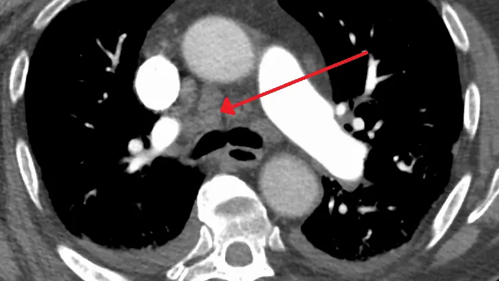
Reading "mediastinal lymphadenopathy" on a chest CT report sounds ominous, partly because "lymph node" and "chest" together make many people think of cancer. The reassuring truth is that it only means one or more lymph nodes in the middle of your chest look larger than average, and the most common reason is the body reacting to an infection or inflammation. Some causes do matter, so it deserves a proper look, but it is a question to work through rather than a diagnosis. Here is what the term means, what usually lies behind it and how the next steps typically go.
What does "mediastinal lymphadenopathy" mean?
The mediastinum is the central compartment of the chest, the space between the two lungs. It holds the heart, the great blood vessels, the windpipe (trachea), the esophagus and a chain of lymph nodes. Lymph nodes are small, bean-shaped filters of the immune system that swell when they are busy, for example while fighting an infection, and "lymphadenopathy" simply means enlarged ones. It is mainly a CT finding: a chest X-ray shows only large groups of nodes, as a widened middle of the chest.
Radiologists measure a node across its short axis, because a normal node is oval and the narrow diameter is the more reliable one. In the mediastinum, a short axis of more than about 1 cm (10 mm) is the usual threshold for "enlarged", with a little more leeway in a few spots such as beneath the fork of the windpipe. A node just over the line may be called "borderline" or "prominent", which says the radiologist noticed it, not that it is a problem.
Reports may also say where the nodes sit, using the lymph node map of the International Association for the Study of Lung Cancer. The labels you are most likely to meet are:
- Paratracheal (stations 2 and 4, with R or L for the side, as in "4R"): alongside the windpipe
- Prevascular (3A): in front of the great vessels
- Aortopulmonary window (5): beneath the arch of the aorta, next to the pulmonary artery
- Subcarinal (7): beneath the point where the windpipe splits in two
- Hilar (10): at the root of each lung. Strictly just outside the mediastinum, but usually reported together with it
What radiologists look at besides size
Size is only the start. A radiologist also weighs shape, inner appearance and the company a node keeps, much as in our guide to an enlarged lymph node on ultrasound or CT:
- Shape and fatty center: an oval node with a small pale fatty center looks like one that is simply reacting. A round node whose fatty center has filled in gets a closer look
- Calcium: dense white flecks usually mean an old, healed infection and are generally harmless. A thin "eggshell" rim of calcium is linked to dust-related lung disease and to sarcoidosis
- A dark center: a low-density middle (necrosis) can come from certain infections, tuberculosis in particular, or from some tumors
- Change over time: nodes unchanged from a scan a year or two ago are reassuring, which is why bringing older images matters
Common causes
Most often a node is reactive: it has enlarged because it is doing its job. The usual explanations are:
- Infection: a pneumonia (see our article on lung consolidation), a heavy chest cold, flu or COVID-19. Nodes can stay enlarged for weeks or months after the illness has gone
- Long-term lung or heart conditions: mild enlargement is common in interstitial lung disease, COPD and heart failure
- Sarcoidosis: an inflammatory condition in which small clusters of immune cells (granulomas) form, most often in the lungs and chest nodes. Nodes at both lung roots and beside the windpipe are the classic picture, and many people have no symptoms
- Tuberculosis and fungal infections: they can enlarge chest nodes and, once healed, leave calcified ones behind. How likely they are depends on where you live and have traveled
Less often, the cause is a disease of the nodes themselves or spread from elsewhere:
- Lymphoma: a cancer of the lymphatic system, for which the middle of the chest is a common site. It may come with fever, night sweats and weight loss, and many types are highly treatable
- Spread from another cancer: lung cancer commonly spreads to the mediastinal nodes, which is why they are checked so closely when a lung nodule is found, as our guide on whether a lung nodule means cancer explains. Breast, kidney and head and neck cancers and melanoma can reach them too
Is it serious?
Usually not. Mild, borderline enlargement is one of the more frequent incidental findings on a chest CT, and after an infection or with a chronic lung condition it is most often reactive and settles on its own. Wording such as "borderline" or "likely reactive" tells you that is how the radiologist read it.
Context decides how seriously it is taken. The same 12 mm node means one thing in someone recovering from pneumonia and another in someone with a known cancer, weight loss and night sweats. Doctors look harder at nodes that are clearly larger than 1 cm, grow on a repeat scan, are round with the fatty center lost, sit at several stations, or come with a lung mass nearby.
Contact your doctor promptly if enlarged nodes come with coughing up blood, trouble swallowing, a new hoarse voice, swelling of the face or neck, worsening breathlessness, or fever with night sweats and weight loss. Interpretation always depends on symptoms, tests and history, not on the scan alone.
Symptoms
Often there are none: the nodes are usually found by chance on a CT done for another reason. When symptoms occur, they come from the cause or, with large nodes, from pressure on nearby structures:
- A cough that will not go away, shortness of breath or chest discomfort
- A hoarse voice or difficulty swallowing, when nodes press on a nerve or the esophagus
- Fever, night sweats, tiredness and weight loss, which point to infection or lymphoma rather than to the nodes themselves
How is it diagnosed and followed up?
The CT is the starting point, ideally with contrast (dye injected into a vein), which helps separate nodes from blood vessels. Comparing with any older scan is often the single most useful step. What follows depends on how the nodes look and on you:
- Mild and low-risk: often a repeat CT after a few months to see whether the nodes have shrunk
- Clinical review and tests: a check for swollen nodes in the neck, armpits and groin, blood tests, tuberculosis tests when risk factors point that way, and breathing tests
- PET-CT: shows which tissue is metabolically busy. It helps stage lymphoma and lung cancer, but infection and sarcoidosis also light up, so a bright node is not proof of cancer. Our guide to hypermetabolic findings and SUV explains the wording
- Biopsy: the only way to know what the cells are. It is often done with EBUS (endobronchial ultrasound), where a thin flexible tube passed through the mouth uses ultrasound to guide a fine needle through the airway wall into a node. Mediastinoscopy (a small cut above the breastbone) is the main alternative
Follow-up recommendations vary by patient, and your own doctor knows your history.
Treatment options
The nodes themselves are not treated; the cause is:
- Reactive nodes: observation, as they shrink once the infection or inflammation settles
- Infection: antibiotics, or antituberculosis or antifungal medicines when those are the cause
- Sarcoidosis: often just monitoring, since many cases settle alone, with steroids or similar medicines if it causes symptoms or affects organs
- Lymphoma: chemotherapy, immunotherapy or targeted drugs, sometimes radiotherapy, chosen by type and stage
- Spread from another cancer: treatment follows the original cancer and is planned by a team
Why a second read can help
A report line about nodes often arrives with little explanation, and the gap between "borderline" and "something more" is where worry grows. Several judgment calls sit inside it. Is an 11 mm node truly enlarged for that spot? Is a pale structure beside the aorta a node, or fluid in a normal fold of the pericardium, which can mimic one? Is a calcified node an old infection, unchanged from the last scan? If you would like an online radiology second opinion, DocOrbit lets you send the study from home, and a subspecialist radiologist writes a structured report you can share with your own doctor. Our guide to when to get a second radiological opinion can help you decide, and the thorax CT second opinion page shows what a full review covers.
Does mediastinal lymphadenopathy mean cancer?
No, not usually. Most mildly enlarged mediastinal nodes are reactive, meaning they have swollen in response to an infection or inflammation, and many shrink on their own. Lymphoma and spread from a lung or other cancer are on the list of possibilities, so the node's features, your history and sometimes a repeat scan or biopsy decide what it is.
What size of mediastinal lymph node is abnormal?
On CT, a node is generally called enlarged when its short axis is more than about 1 cm. Size alone cannot separate harmless from significant, though: a 12 mm reactive node and a 12 mm cancerous one can look alike, so shape, fatty center, calcium and change over time count as well.
Do I need a biopsy for enlarged mediastinal lymph nodes?
Not always. A biopsy is usually kept for nodes that are clearly enlarged, growing or suspicious on PET-CT, or when the answer would change treatment. If one is needed, a procedure called EBUS can often sample the nodes through the airways without an operation.
Key takeaways
- Mediastinal lymphadenopathy means enlarged lymph nodes in the middle of the chest, usually a short axis above about 1 cm on CT
- Most mildly enlarged nodes are reactive to an infection or inflammation, and many shrink on their own
- Shape, fatty center, calcium, change over time and your history matter more than size alone
- Coughing up blood, trouble swallowing, facial swelling or fever with night sweats and weight loss need prompt attention
If the rest of your report reads like another language, our free radiology report explainer rewrites it in everyday words. And if you would like to go through the images with a specialist, you can ask a radiologist online through DocOrbit and take the answers back to your own doctor.
Can I get a second opinion on my chest CT scan online?
Yes. You upload your images from home, with no CD to post and no second trip, and a subspecialist radiologist re-reads the study and writes a structured report you can hand to your own doctor within 24 to 48 hours. You can message the radiologist with questions about the scan afterwards, including what the wording about your lymph nodes means for you.
This article is for general information only and is not medical advice. Always discuss your imaging results and any next steps with a qualified physician.