
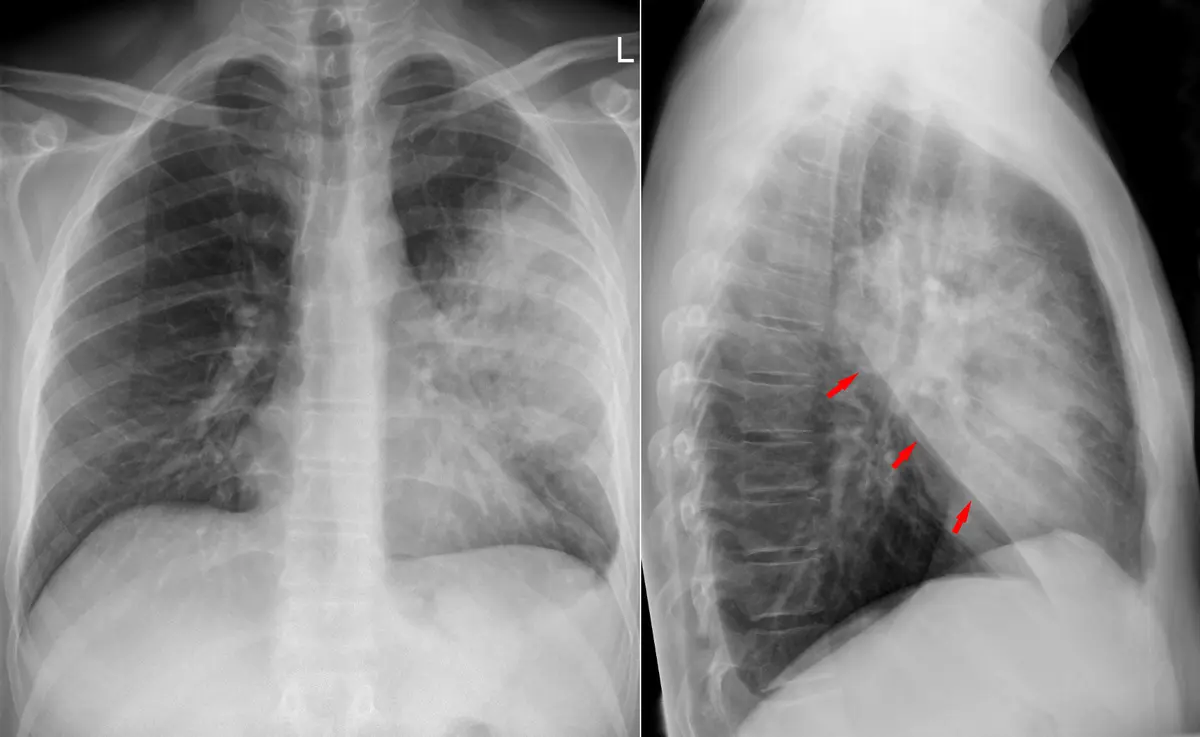
Seeing the word "consolidation" on a chest X-ray or CT report can be unsettling, partly because it sounds hard and permanent. The reassuring truth is that it describes how part of the lung looks, not a diagnosis, and the most common cause is an infection such as pneumonia, which usually responds well to treatment. Here is what it means, what causes it and what happens next.
What does "consolidation" mean?
Your lungs are made of millions of tiny air sacs, called alveoli. Healthy lung is mostly air, so it looks dark on a chest X-ray and CT. In consolidation, the air in one area has been replaced by something else, most often pus or fluid and sometimes blood or inflammatory cells, so that patch turns white and solid-looking. The Fleischner Society's glossary of thoracic imaging terms, the standard reference, describes it the same way.
It can involve a small patch, a whole lobe (one of the five main sections of the lungs) or several areas at once. Radiologists look for a few things:
- On a chest X-ray, a white, fairly even patch with fuzzy edges. It may follow the shape of a lobe and stop sharply at a fissure, the thin line between lobes. Where it touches the heart or diaphragm, that outline fades, which helps place it
- Air bronchograms: dark, branching lines inside the white patch. They are airways that still hold air while the sacs around them are filled
- On CT, the patch is dense enough to hide the vessels running through it, unlike ground-glass opacity, where they stay visible
Consolidation usually does not shrink the lung, which helps tell it apart from atelectasis, where part of the lung deflates and loses volume. The two can occur together.
Common causes
Consolidation is usually grouped by what has filled the air sacs:
- Pus from an infection: pneumonia is by far the most common cause, whether bacterial, viral or from inhaled food or stomach contents (aspiration)
- Fluid: fluid backing up when the heart is not pumping well (pulmonary edema), usually in both lungs
- Blood: bleeding into the lung after a chest injury or from some blood-vessel or autoimmune conditions, or a wedge-shaped area after a blood clot in the lung
- Inflammation and other causes: organizing pneumonia (a slow-healing inflammation of the air sacs), reactions to some medicines, chest radiation and, uncommonly, certain cancers that grow along the air sacs and mimic a pneumonia that does not clear
Fluid can also collect around the lung in pneumonia, and our guide to pleural effusion explains how that is found and treated.
Is it serious?
That depends on the cause and on who the patient is. Because pneumonia is the usual explanation, consolidation is most often treatable: many otherwise healthy adults recover at home with antibiotics, rest and fluids, while older adults, babies and people with heart or lung disease or weakened immunity are watched more closely and sometimes treated in hospital.
Doctors look harder when a consolidation does not clear after treatment, returns in the same place or appears in a long-term smoker or an older person. The question then is whether something is blocking an airway or growing in the lung, such as a tumor, which is far from the usual explanation. If your report described a round spot rather than a hazy patch, see our guide to lung nodules and cancer.
Breathlessness at rest, confusion, bluish lips, worsening chest pain, coughing up blood or a high fever that will not settle need prompt medical attention. As with most imaging findings, interpretation depends on your symptoms, examination, tests and history, not on the scan alone.
Symptoms
When the cause is pneumonia, the usual picture is:
- A cough, dry at first and often later with phlegm
- Fever, chills and sweating
- Shortness of breath or fast breathing
- Chest pain that is worse when you breathe in deeply or cough
- Tiredness and loss of appetite
Older adults may only seem confused or unusually tired, and consolidation is sometimes found by chance on an X-ray taken for another reason.
How is it diagnosed and followed up?
A chest X-ray is usually the first test. A CT is added when the X-ray is unclear, when you are not improving as expected, when a complication such as fluid around the lung or an abscess (a pocket of pus) is suspected, or to look for an underlying cause. The image is read together with your symptoms, examination, oxygen level and often blood tests. If a blocked airway is suspected, a doctor may recommend bronchoscopy, in which a thin, flexible camera looks inside the airways.
The picture clears more slowly than you feel better: the white patch can take several weeks to fade, longer in older people and smokers, even after successful treatment. Many people who recover fully need no repeat image, while an X-ray or CT after a few weeks is commonly suggested if symptoms linger or the risk of an underlying lung problem is higher. Follow-up recommendations vary by patient, and your own doctor knows your history.
Treatment options
Treatment targets the cause, not the white patch itself:
- Bacterial pneumonia: antibiotics, plus rest, fluids and medicine for fever and pain, with hospital care and oxygen if severe
- Viral pneumonia: mostly rest and fluids, sometimes an antiviral medicine
- Fluid from the heart: medicines that remove the extra fluid, and treatment of the heart condition behind it
- Inflammatory causes: such as organizing pneumonia, often treated with corticosteroids (anti-inflammatory medicine)
- A tumor: uncommon, and leads to referral for further tests
Why a second read can help
"Consolidation" is a description, and what it means for you depends on details that are easy to pass over: which lobe is involved, whether fluid sits beside it, whether an airway looks blocked, and whether something else, such as a small nodule, hides behind it or has changed since an earlier scan. A thoracic radiologist reads exactly these details every day. If you would like a radiology second opinion online, DocOrbit lets you send your scan from home, and a subspecialist radiologist writes a structured report you can share with your own doctor. Our guide on when to get a second radiological opinion can help you decide, and the thorax CT second opinion page shows what a full chest CT review covers.
Is consolidation the same as pneumonia?
Not exactly. Consolidation describes how part of the lung looks on a scan, with the air replaced by fluid, pus, blood or cells. Pneumonia is a diagnosis your doctor makes by combining the image with your symptoms, an examination and tests. Most consolidation is pneumonia, but not all of it.
How long does consolidation take to clear on a chest X-ray?
Usually several weeks, and the image often lags behind how you feel. The white patch can still be visible after the fever and cough have gone, and it fades more slowly in older people, smokers and people with lung disease. Your doctor decides whether a repeat image is needed, which is more likely if symptoms linger.
Can consolidation be a sign of lung cancer?
Occasionally, but it is far from the usual explanation, since most consolidation is infection. Certain cancers can fill the air sacs and look like pneumonia that does not clear, so a consolidation that persists after treatment or returns in the same place usually prompts a CT or a look inside the airways. That is a thorough workup, not a diagnosis.
Key takeaways
- Consolidation means air in part of a lung has been replaced by pus, fluid, blood or cells. It describes how the lung looks, not a diagnosis
- Pneumonia is by far the most common cause, and it usually responds well to treatment
- The white patch can take weeks to clear, even after you feel better
- One that persists or returns in the same place is looked at more closely
If the rest of your report reads like another language, our free radiology report explainer rewrites it in everyday words. And if you would rather go through the images with a specialist, you can ask a radiologist online through DocOrbit and take the answers back to your own doctor.
How do I have my chest scan reviewed by another radiologist online?
You upload your images from home, with no CD to post and no extra trip, and a subspecialist radiologist re-reads the study and writes a structured report you can hand to your own doctor within 24 to 48 hours. You can message the radiologist with questions about the scan afterwards, including what the wording on your report means for you.
This article is for general information only and is not medical advice. Always discuss your imaging results and any next steps with a qualified physician.
