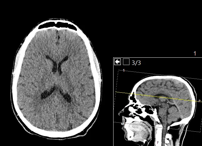
You already have the scan. Maybe you went to the emergency room after a fall or a sudden, severe headache, and the report came back reassuring but something still does not sit right. Maybe it flagged a finding you do not fully understand, or you simply want your head CT read again, closely, by someone who is not working through a stack of other cases at once. A head CT second opinion, sometimes called a brain CT second opinion, is exactly that: a board-certified neuroradiologist reviews your existing images independently and sends back a written report you can bring straight to your own doctor. Here is what that second read involves, why this scan rewards the extra attention, and what happens after you send it in.
A head CT is read fast, because it has to be
A head CT is usually ordered because something needs an answer quickly: a fall, a sudden severe headache, new confusion, weakness on one side, a possible stroke. The scan itself takes only a few minutes, and that speed is the whole point in an emergency, which is also why the first read is built for speed. In a busy emergency department, or an overnight teleradiology shift covering several hospitals at once, a head CT is often read within minutes, because the question in front of the radiologist is urgent: is there bleeding, is there a large stroke, is there dangerous swelling on the brain.
That kind of read is very good at catching what it was built to catch. It is less suited to a small, quiet finding that was not the reason the scan was ordered, an early lacunar infarct tucked in the basal ganglia, a small meningioma against the falx, a patch of chronic small-vessel change, some mucosal thickening in a sinus at the edge of the image. None of this reflects badly on the radiologist who read it fast under real pressure, it reflects what volume and urgency do to any read. A slower, unhurried second look at the same images buys back the attention the first pass did not have time for.
Why a neuroradiologist reads a head CT differently
A general radiologist reads everything that comes through the department, chest X-rays, abdominal CTs, mammograms, and head CTs, often within the same shift. A neuroradiologist reads brain and spine imaging exclusively, often for years, and that narrow focus changes what stands out. Someone who reads head CTs constantly is quicker to catch a few millimeters of blood layered along the tentorium that could pass for normal density, a loss of the grey-white distinction marking a very early stroke, or a subtle shift of the midline structures that is easy to miss without a careful comparison.
This is the logic behind asking a subspecialist for a head CT second read rather than a general reader: not that the first radiologist was careless, but that a narrower field of view statistically catches patterns a broad one is more likely to under-call. Spine and head and neck imaging round out the same subspecialty, which is why a neuroradiologist also reads a scan that extends to the skull base or upper neck.
What a head CT actually shows, structure by structure
A thorough head CT review works through the same anatomy a neuroradiologist checks on every study, structure by structure:
- The skull and scalp — checked for fractures, from a simple linear crack to a depressed fracture or one at the skull base, and for scalp swelling that can point to where an impact happened
- The extra-axial spaces, the thin spaces between the skull and the brain — an epidural hematoma (a lens-shaped collection, typically from a torn artery), a subdural hematoma or hygroma (a crescent-shaped collection along the brain's surface), and subarachnoid hemorrhage (blood in the grooves and cisterns) are all assessed here
- The brain tissue itself — reviewed for a bleed, for the density changes of an acute or evolving stroke, for chronic ischemic small-vessel changes, and for any mass effect or shift of the normally symmetric midline structures
- The ventricles and CSF spaces — their size and shape can point toward hydrocephalus, or reflect ordinary age-related cerebral atrophy, told apart by pattern, not size alone
- The basal cisterns, the fluid-filled spaces around the brainstem — checked for effacement, a sign that pressure inside the skull may be rising, and for blood layering within them
- Calcifications — the pineal gland, choroid plexus, and falx normally calcify with age, so a careful read separates that from calcification inside a mass such as a meningioma
- The paranasal sinuses and mastoids — almost always partly visible on a head CT, commonly showing incidental mucosal thickening
- The skull base and visible bone marrow — reviewed on its own for fractures, separate from the brain tissue it surrounds
Two honest limits are worth stating plainly. A plain head CT has less soft-tissue detail than an MRI, so a small mass or a stroke only a few hours old can be easier to miss on CT, and a careful second read says so rather than promising more than the images show. And a non-contrast head CT is not an angiogram: a dense clot inside a major vessel can sometimes be suspected, but confirming or ruling out a brain aneurysm or a narrowed vessel needs a dedicated CT angiogram (CTA), a separate, contrast-based study built to show the vessels. A trustworthy head CT interpretation draws that line clearly.
How DocOrbit reads your head CT
Send us the same images, the DICOM files from the scan or a copy of the disc you were given, and a board-certified neuroradiologist performs a full head CT second read, going through the same structures one at a time, from the scalp and skull to the extra-axial spaces to the skull base, and writes a structured report you can hand directly to your own doctor. This is an online radiology second opinion built around images you already have, no new scan needed. The signed report comes back in 24–48 hours, and it is not a verdict on the first read being wrong, only what a fresh, focused review of your head CT found, in the clinical language your physician already speaks.
Get your head CT re-read by a subspecialist
Ask the radiologist questions afterward
One thing that surprises most patients: the second opinion does not end when the report lands in your inbox. Once you have it, you can message the radiologist directly, about a phrase in the report or what a finding means for you, and get a written answer back. Most people never get the chance to ask the person who actually looked at their images anything at all.
Or get an answer in minutes with Orbius
If waiting 24–48 hours is not what you need right now, DocOrbit also offers Orbius, an AI radiology agent that reads the same head CT and returns a structured report, findings, impression, next steps, in minutes rather than days, at a lower cost, any time of day. Orbius is the fastest way to upload your scan for a second opinion when a signed report does not fit your timeline, an honest trade of speed against a subspecialist's training and signature. Upgrading to a full neuroradiologist read from the same upload is one click away.
How do I send my head CT for a second opinion?
Upload the DICOM files from your scan, or the CD you were given at the imaging center or emergency room, directly through the DocOrbit platform. You don't need to mail anything or visit an office, the images go straight to the radiologist reading your case.
Do I need the actual CT images, or is the written report enough?
You need the images themselves, not just the written report. A genuine second opinion means a radiologist looks at your head CT directly, the same way the first one did, rather than reviewing someone else's description of what they saw.
Will my own doctor accept a second opinion report?
Yes. The report follows the same structured format radiologists use everywhere, with findings and an impression your doctor can read and act on like any other radiology report, and you are free to share it with them directly.
How long does a head CT second opinion take?
A signed report from a board-certified neuroradiologist typically comes back in 24 to 48 hours. If you want an answer sooner, Orbius, DocOrbit's AI radiology agent, returns a structured report on the same images in minutes.
Is it worth getting a second opinion if my head CT was reported as normal?
It can be, especially if your symptoms have not improved or do not match the normal label, or if the scan was read quickly for one urgent question that was not actually yours. A fresh, unhurried head CT review sometimes turns up something the original fast read was not looking for, and just as often confirms that normal really does mean normal.
Key takeaways
- A head CT produces dozens of thin slices, and the clinical question behind it is usually urgent, so the first read is built for speed
- A neuroradiologist reads brain and spine imaging exclusively, and catches patterns a generalist is more likely to under-call
- A proper second opinion walks the same anatomy structure by structure, and says plainly when a finding needs CTA for the vessels or MRI for soft-tissue detail
- DocOrbit's signed neuroradiologist read takes 24–48 hours; Orbius, the AI option, returns a structured report in minutes
- You can message the radiologist with questions after the report arrives
Can I get a second opinion on my CT scan online?
Yes. You upload your images from home, with no CD to post and no second trip, and a subspecialist radiologist re-reads the study and writes a structured report you can hand to your own doctor within 24 to 48 hours. You can message the radiologist with questions afterward, and Orbius can return a first read in minutes if you want an answer sooner.
This article is for general information only and is not medical advice. Always discuss your imaging results and any next steps with a qualified physician.
