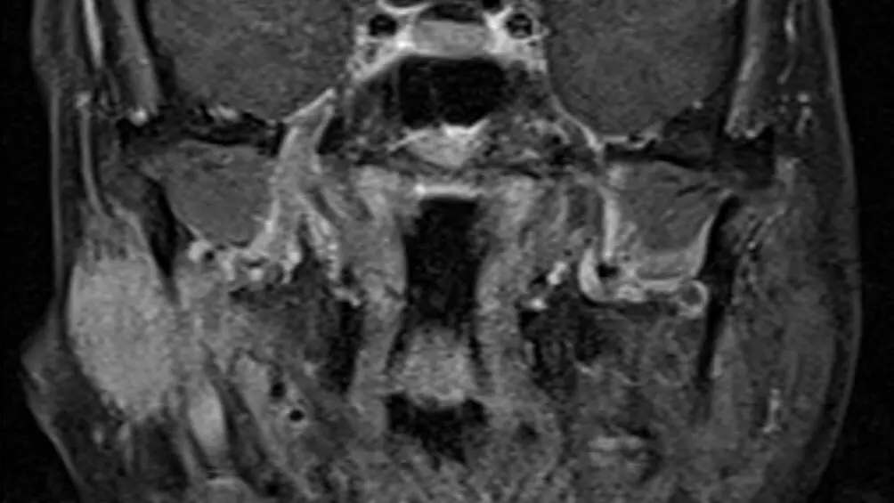
You already have the scan. Maybe the report came back reassuring and something still does not sit right, maybe it describes a lymph node, a gland or a skull base finding you do not fully understand, or maybe you are about to see an ear, nose and throat (ENT) surgeon and want your head and neck MRI re-read by someone who looks at this region every day. A head and neck MRI second opinion is exactly that: a board-certified neuroradiologist reviews your existing images independently and sends back a written report you can bring straight to your own doctor. Here is what that second read involves and what happens after you send it in.
A head and neck MRI packs a lot into a small space
A head and neck MRI covers everything from the skull base to the collarbones: the face, nose and sinuses, mouth and throat, salivary glands, the soft tissues and lymph nodes of the neck, and often the lower edge of the brain. It runs several sequences, T1, fat-suppressed T2, diffusion and usually contrast-enhanced series, in more than one plane, so one study can run to hundreds of images, and almost every structure sits within a few centimeters of the next, separated by thin planes of fat that a tumor, an infection or a nerve can cross quietly.
In a busy department, a general radiologist works through that stack alongside chest, abdomen and spine studies, and the read is shaped by the question on the request form: is this lump a gland or a node, has this known tumor grown. The rest of the field of view gets less attention, and that is where small findings live: a sub-centimeter node at the end of a chain, a quiet change at a skull base opening, a thin line of enhancement along a nerve. None of this is a comment on any radiologist's competence; it is what volume and time do to any read, and a slower second read gives the attention back.
Why a neuroradiologist reads a head and neck MRI differently
A general radiologist reads every body part in the same shift. Head and neck imaging sits within neuroradiology, and a neuroradiologist who reads this region regularly has seen thousands of variations that look alarming and are not, and the smaller number that look ordinary and are not. They are quicker to notice that one side of the nasopharynx is subtly fuller than the other, that a lymph node has lost its normal fatty center, that enhancement along a cranial nerve is a thread of tumor rather than an ordinary vessel, or that a parotid nodule sits in the deep lobe rather than the superficial one, which changes what a surgeon plans.
That is the logic behind asking a subspecialist for a head and neck MRI second read: not that the first radiologist was careless, but that a narrower, deeper field of view catches patterns a broad one is more likely to under-call.
What a head and neck MRI shows, structure by structure
A thorough head and neck MRI interpretation works through the same compartments on every study:
- The nasopharynx and skull base: where nasopharyngeal carcinoma arises, plus the bone and marrow of the skull base around it, checked for tumor spread, marrow replacement and rarer growths such as chordoma
- The mouth, tongue and oropharynx: the tonsils, tongue base and floor of the mouth, assessed for masses, how deep they reach and whether the jaw marrow is involved; dental work is safe in the scanner (see having an MRI with metal in your body), though fillings and braces can blur this area
- The salivary glands: the parotid and submandibular glands, for benign tumors such as pleomorphic adenoma and Warthin tumor, for malignant ones, and for inflammation
- Lymph nodes and the deep neck spaces: node chains assessed for size, shape, a necrotic center and spread beyond the capsule (see enlarged lymph nodes on ultrasound and CT), plus the carotid space (paraganglioma, nerve sheath tumors) and the retropharyngeal and parapharyngeal spaces
- The cranial nerves: the trigeminal and facial nerves and the skull base openings they pass through are followed for thickening or enhancement, because some tumors travel along a nerve, far from the main mass; MRI is the preferred test for this perineural spread, and it is easy to miss if no one is looking
- The orbits and optic nerves: optic neuritis, optic nerve sheath meningioma, thyroid eye disease with enlarged eye muscles, and masses of the orbit or tear gland
- The paranasal sinuses and nasal cavity: mucosal thickening, polyps and retention cysts, and whether something filling a sinus is trapped secretions or a tumor, which MRI separates better than CT
A few honest limits are worth stating, because a good report states them too. Fine bone detail, such as thin sinus walls, is shown better by CT, and the larynx is usually assessed with endoscopy and CT because swallowing and breathing blur it on MRI, though MRI can add detail about cartilage and spread in a known tumor. Ultrasound is the test that characterizes a thyroid nodule (see thyroid nodules on ultrasound). Blood vessels show only as dark flow voids, so judging a narrowed or torn carotid artery or a clotted vein takes an MR angiogram (MRA), CT angiogram or Doppler ultrasound, and for a known cancer PET-CT is often added to look for spread elsewhere. The lower brain and pituitary region sit at the margin of the frame and have their own protocol, as in our brain MRI second opinion article.
How DocOrbit reads your head and neck MRI
Send us the same images, the DICOM files from the scan, and a board-certified neuroradiologist subspecialized in head and neck imaging performs a full second read, going through the same compartments one at a time, from the skull base and nasopharynx through the glands and nodes to the orbits and sinuses, and writes a structured report you can hand directly to your ENT surgeon, oncologist or family physician. It is a radiology second opinion online, built around images you already have: no new scan, no new appointment. The signed report comes back in 24–48 hours, and it is not a verdict on the first read, only what a fresh, focused second read of your head and neck MRI found. To have it re-read, upload your scan for a second opinion below.
Get your head and neck MRI re-read by a subspecialist
Ask the radiologist questions afterward
One thing that surprises most patients: the second opinion does not end when the report lands in your inbox. You can message the radiologist directly with questions, about a phrase in the report, or about what a finding does or does not mean for you, and get a written answer back. You can ask a radiologist online why the report says what it says about a node or a gland, which most people never get to do with the person who actually looked at their images.
Or get an answer in minutes with Orbius
If waiting 24–48 hours for a signed report is not what you need right now, DocOrbit also offers Orbius, an AI radiology agent that reads the same head and neck MRI and returns a structured report with findings, a conclusion and next steps in minutes, at a lower cost, any time of day. Orbius is the route to MRI report interpretation online when time matters more than a signature, an honest trade of speed and price against a human subspecialist's training. If its report raises something you want a specialist to weigh in on, upgrading to a full neuroradiologist read from the same upload is one click away.
How do I send my head and neck MRI for a second opinion?
Upload the DICOM files from your scan, or the CD you were given at the imaging center, directly through the DocOrbit platform. You do not need to mail anything or visit an office, the images go straight to the radiologist reading your case.
Do I need the actual MRI images, or is the written report enough?
You need the images themselves, not just the written report, and the complete study, including the contrast-enhanced and diffusion series. A genuine second opinion means a radiologist looks at your head and neck MRI directly, the way the first one did, rather than reviewing someone else's description of it.
Will my own doctor accept a second opinion report?
Yes. The report follows the same structured format radiologists use everywhere, with findings and an impression that your ENT specialist, oncologist or family doctor can act on like any other radiology report, and you are free to share it with them directly.
How long does a head and neck MRI second opinion take?
A signed report from a board-certified neuroradiologist typically comes back in 24 to 48 hours. If you want an answer sooner, Orbius, DocOrbit's AI radiology agent, returns a structured report on the same images in minutes.
Is it worth getting a second opinion if my head and neck MRI was reported as normal?
It can be, especially if symptoms such as a lump, hoarseness or facial numbness have not settled, or if the scan was read quickly for one specific question that was not actually yours. A fresh, unhurried head and neck MRI review sometimes finds something the original read was not looking for, and just as often confirms that normal really does mean normal.
Key takeaways
- A head and neck MRI packs the face, throat, glands, nodes, nerves and skull base into hundreds of images, more than one fast read is built to comb fully
- A neuroradiologist reads this region regularly and catches patterns, such as perineural spread, that a generalist is more likely to under-call
- A proper second opinion goes through the same compartments one by one and says plainly when a question belongs to CT, ultrasound or endoscopy
- DocOrbit's signed read takes 24–48 hours; Orbius, the AI option, returns a structured report in minutes
- You can message the radiologist with questions after the report arrives
Can I ask a radiologist about my MRI report online?
Yes. You upload your images from home, with no CD to post and no second trip, and a subspecialist radiologist re-reads the study and writes a structured report you can hand to your own doctor within 24 to 48 hours. You can message the radiologist with questions afterward, and Orbius can return a first read in minutes if you want an answer sooner.
This article is for general information only and is not medical advice. Always discuss your imaging results and any next steps with a qualified physician.
