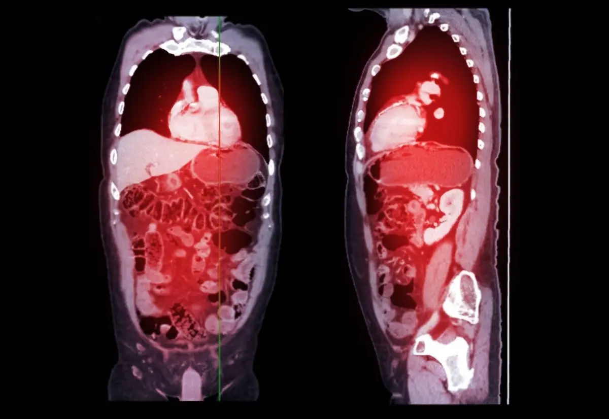
If your CT report mentions "fat stranding," "heterogeneity of the fat planes," or "increased density in the surrounding fat," it is describing the appearance of the body's fat rather than naming a disease. Normally fat looks smooth and uniformly dark on a scan; when something nearby irritates it, thin hazy lines appear in it. That is all the phrase means. It is one of the most common observations in radiology, it is usually caused by something benign and temporary, and it is almost never a diagnosis on its own. This article explains what the radiologist is actually seeing, what tends to cause it, and when it changes the plan.
What does "fat stranding" mean?
Fat is an unusually helpful background on a CT scan. It has a very low density, so on the images it appears almost uniformly black, and it fills the spaces between organs, muscles, and blood vessels. Against that flat dark backdrop, even a small change shows up clearly.
When fluid, blood, inflammatory cells, or congested lymphatic channels seep into a patch of fat, its density rises slightly and it stops looking uniform. On the images this shows up as fine grey lines running through the black, like faint spider-web threads or wisps of smoke. Radiologists call those lines "strands," and the fat itself gets described in a handful of interchangeable ways:
- Fat stranding or stranding of the adjacent fat — the standard phrase
- Dirty fat or misty fat — informal, and no comment on hygiene
- Haziness, reticulation, or increased attenuation of the fat — more technical descriptions of the same texture
- Fat plane heterogeneity — the fat is no longer uniform in appearance, often used on MRI and ultrasound reports
- Blurring or loss of the fat planes — the clean fat line that normally separates two structures is harder to make out
The finding is not limited to CT. On MRI the same process shows up as increased signal in the fat on fluid-sensitive sequences, and on ultrasound as fat that looks brighter and more echogenic than it should. CT is simply where the wording is most familiar, because fat contrasts so strongly with everything around it.
Why radiologists pay attention to it
Fat stranding is valuable less for what it is than for where it is. Because fat reacts to whatever sits next to it, the haziness usually surrounds the actual problem, working like an arrow pointing at the organ involved.
Stranding wrapped around the appendix supports appendicitis. Stranding hugging a segment of colon suggests diverticulitis. Stranding around the pancreas raises the question of pancreatitis, around the gallbladder cholecystitis, and in the fat next to a kidney it can accompany an obstructing kidney stone. In several of these conditions the organ itself can look nearly normal in the early hours, and the reaction in the fat is the first visible clue that anything is wrong.
That sensitivity is also the catch. A sign that responds to almost anything is, by definition, not specific to any one thing.
Common causes
The list is long, and most of it is unremarkable:
- Inflammation or infection of a neighboring organ — appendicitis, diverticulitis, pancreatitis, cholecystitis, and their equivalents elsewhere in the body
- Recent surgery, biopsy, or drain placement — stranding at an operated site is expected and can linger for weeks
- Trauma or bruising, including minor injuries you may not remember
- Fluid overload from heart, kidney, or liver conditions, or low blood protein levels, which typically produces widespread rather than focal haziness
- Venous or lymphatic congestion, where drainage from an area is partly blocked
- Passage of a stone through the ureter or bile duct, which irritates the fat along the way
- Previous radiotherapy to the region
- Self-limiting conditions of the fat itself, such as epiploic appendagitis or omental infarction, which mimic more serious abdominal emergencies and resolve on their own
- Mesenteric panniculitis, a chronic and usually harmless hazy appearance of the fat that anchors the small bowel
- Tumor involvement, when a cancer irritates or extends into the surrounding fat — real, but well down the list of frequencies
Is fat stranding serious?
That depends far more on the pattern and your symptoms than on the words themselves. Three features shape the interpretation.
Focal versus diffuse. A tight, well-defined patch of stranding centered on one organ is a meaningful localizing sign. Faint haziness spread across the whole abdomen or under the skin usually points to body-wide fluid balance rather than any single disease.
How intense it is. A few wispy lines are a much softer finding than dense, confluent stranding with free fluid, thickened tissue planes, or gas bubbles alongside it.
What you have been feeling. Focal stranding in someone with sharp right lower abdominal pain, fever, and a raised white cell count is treated urgently. The identical appearance in someone with no symptoms, scanned for an unrelated reason, is often noted and left alone.
Reports frequently pair the finding with hedging language such as "nonspecific," "may reflect inflammatory change," or "correlate clinically." That is not evasiveness. It is an honest acknowledgment that this particular appearance genuinely cannot be pinned to one cause from the images alone.
What "loss of fat planes" adds
A related phrase deserves its own note, because it worries people more than it usually should. The body keeps thin sheets of fat between adjacent structures, and those sheets are easy to see on a scan. When a report says the fat plane between two structures is "lost," "effaced," or "no longer clearly seen," it means that visible dividing line has disappeared.
One explanation is that something is growing across the boundary. Several far more common ones are that swelling has blurred it, that previous surgery or radiation has scarred it, that the person is naturally slim and has little fat there to begin with, or that the imaging plane simply cut across the boundary at an awkward angle. When invasion is genuinely in question, radiologists look for supporting evidence such as an irregular interface, distorted anatomy, or abnormal lymph nodes, and often ask for a dedicated study with contrast before drawing any conclusion.
What usually happens next
Follow-up is driven by the suspected cause rather than by the stranding itself. In an acute setting the finding is combined with your examination and blood tests, and treatment for the underlying condition typically begins the same day. For a self-limiting condition such as epiploic appendagitis, the answer is often nothing more than pain relief and time.
When stranding turns up incidentally in someone with no symptoms, the usual course is either no action at all or a repeat scan after an interval, on the reasoning that a reactive change should settle while something that matters will not. If it persists unchanged, further characterization may be suggested, particularly around organs where it can accompany other findings, such as the pancreas.
Why a second read can help
Fat stranding is one of those findings where the value lies almost entirely in the interpretation. Whether the haziness is focal or diffuse, whether it is centered on one organ or drifting across several, and whether it fits the symptoms that brought you in are all judgment calls made by the reader, and reasonable radiologists sometimes weigh them differently. DocOrbit offers an expert second read of your scan that you can share directly with your own doctor, which people often find useful when a single descriptive line has changed the plan. If that sounds relevant, our guide to when to get a second radiological opinion and our broader look at the role of second opinions in radiology are good starting points.
Does fat stranding always mean infection?
No. Infection is only one of the things that makes fat look hazy, and plenty of causes have nothing to do with germs. Recent surgery, a biopsy, an injury, a blocked vein, fluid retention from heart or kidney problems, and simple irritation from a passing stone all produce the same appearance. The radiologist works out which of those fits by looking at where the stranding is, how far it spreads, and what your symptoms and blood tests show.
Can fat stranding go away?
Yes, and it usually does once whatever caused it settles down. Fat stranding is a reaction rather than permanent damage, so it fades as inflammation resolves, a stone passes, an operation heals, or excess body fluid is cleared. Resolution on a follow-up scan is one of the ways doctors confirm that treatment worked, though the fat can lag a week or two behind how you feel.
What does loss of fat planes mean in a report?
It means the thin layer of fat that normally separates two structures can no longer be seen as a clean line between them. That can happen because something is growing across the boundary, but it happens far more often because swelling, inflammation, previous surgery, or simply a slim body build has blurred the border. On its own the phrase is a description of the picture, not a statement that anything has spread.
Does fat stranding mean cancer?
In the great majority of scans it does not. The common causes are inflammatory, post-surgical, or related to fluid balance, and cancer is well down the list. Tumors can irritate or seed the surrounding fat, which is why a radiologist reads the stranding alongside the presence of a mass, enlarged lymph nodes, and your history rather than treating the haziness as a finding by itself.
Why does my report mention fat stranding when I feel fine?
Mild or widespread haziness in fat is a common incidental observation on scans done for other reasons, and it often reflects nothing more than fluid balance, previous surgery, or a change that has already healed. Radiologists describe what they see, so the phrase appears in the report even when it carries no clinical weight. Focal stranding in someone with matching pain is a very different situation from diffuse stranding in someone with no symptoms at all.
Key takeaways
- Fat stranding describes hazy grey lines in fat that normally looks uniformly dark, and it is a reaction to something nearby rather than a diagnosis
- Its main value is as a pointer: the fat next to an irritated organ reacts first, sometimes before the organ itself looks abnormal
- Common causes are inflammation, recent surgery, injury, stones, and fluid overload, with tumor involvement much further down the list
- Focal stranding matching your symptoms carries real weight, while faint diffuse haziness in someone who feels well usually does not
- Loss of fat planes is a description of a blurred border, most often from swelling or scarring rather than from anything spreading
- The finding typically fades once the underlying cause resolves, which is why a repeat scan is sometimes all that is needed
This article is for general information only and is not medical advice. Always discuss your imaging results and any next steps with a qualified physician.