
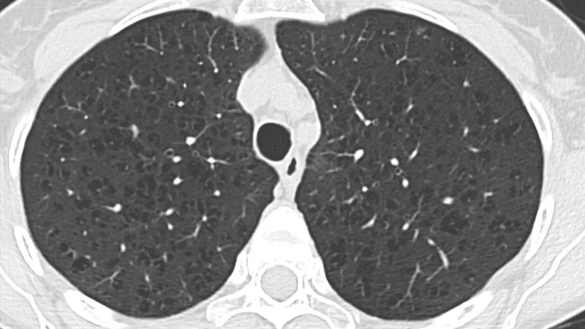
Reading "emphysema" or "air trapping" on a chest CT report can be frightening, because both sound like the end stage of a long illness. The reassuring truth is that mild emphysema is common, often found by chance, and describes how the lung tissue looks rather than how well you breathe. It is a lasting change, so it deserves attention, but a great deal can be done to keep it from progressing.
What do "emphysema" and "air trapping" mean?
Your lungs end in millions of tiny air sacs, the alveoli, whose thin walls pass oxygen into the blood. In emphysema those walls break down, and neighboring sacs merge into larger, floppier air spaces. The lung loses its springiness, which makes it harder to push air out, and it has less surface for oxygen to cross. Tissue that is lost does not grow back.
Air trapping is a different observation. When you breathe out, healthy lung empties and looks denser on CT. In air trapping, part of the lung stays inflated because the small airways leading to it are narrowed or blocked, so air gets in more easily than it gets out.
On imaging, radiologists look for a few things:
- On CT, emphysema appears as patches of very dark lung, as if punched with tiny holes that have no visible wall. Radiologists name it by pattern: centrilobular (small holes, mostly in the upper lungs, the typical smoking pattern), panlobular (spread evenly, more at the bottom, linked to a rare inherited cause) and paraseptal (along the edges of the lung). A hole larger than about 1 cm is called a bulla
- On a chest X-ray, usually only moderate or severe emphysema shows: lungs that look overinflated, flattened diaphragms and fewer visible vessels. Mild emphysema is easily missed, which is why CT is the better test
- Air trapping is seen on scans taken while you breathe out, where the trapped area stays dark while healthy lung turns whiter. A patchy, mosaic look on the ordinary breathing-in pictures, called mosaic attenuation, is a clue that sends the radiologist looking for it
A small amount of air trapping, especially in the lower lungs, is common even in healthy people and is not usually a concern. Report wording such as "mild", "scattered" or "upper-lobe predominant" describes how much there is and where.
Common causes
The two findings have different usual suspects. For emphysema:
- Smoking: by far the most common cause. Heavy dust or fumes at work, or smoke from cooking and heating with wood or other solid fuels, add to it
- Alpha-1 antitrypsin deficiency: an inherited shortage of a protein that protects the lungs. It can cause emphysema in younger adults, including people who never smoked, and often lies behind the panlobular pattern
For air trapping, the cause is usually something that narrows the small airways:
- Asthma and COPD (chronic obstructive pulmonary disease, the umbrella term for long-term airflow limitation)
- Bronchiolitis: inflammation of the smallest airways, often after an infection
- Hypersensitivity pneumonitis: an allergic-type inflammation from breathing in certain dusts, such as mold or bird proteins
- A partly blocked airway: a mucus plug, an inhaled object or, less often, a growth can let air in but not out
Inflamed small airways also produce the pattern described in our guide to tree-in-bud on a chest CT. And a blockage that lets no air in at all leads to the deflated area covered in our article on atelectasis.
Is it serious?
It depends on how much lung is affected and, above all, on how you breathe. Mild emphysema covering a small part of the lungs often causes no symptoms and no measurable loss of lung function. It is frequently reported as an incidental finding, for example on a CT done to screen for lung cancer in people with a long smoking history. Extensive emphysema is different: it is a main component of COPD and can cause lasting breathlessness. The change itself is permanent, but its course is not fixed, and stopping smoking, where it applies, slows it more than any other single step.
Emphysema on a CT is not the same as a diagnosis of COPD, which rests on a breathing test. Some people with emphysema on CT blow normally, so your doctor reads the scan together with your breathing test and symptoms.
Emphysema is also linked to a somewhat higher chance of lung cancer, on top of the risk from smoking itself, which is one reason radiologists look closely at any nodule in emphysematous lung. Most nodules are still benign, as our guide on whether a lung nodule means cancer explains.
Sudden chest pain with shortness of breath (a bulla can rupture and let air leak around the lung), coughing up blood, breathlessness that worsens quickly, or a fever with more or darker phlegm than usual all need prompt medical attention. As with most imaging findings, interpretation depends on your symptoms, breathing tests and history, not the scan alone.
Symptoms
Mild emphysema and small amounts of air trapping often cause nothing at all. When emphysema is more extensive, the usual picture builds slowly:
- Shortness of breath, first on stairs or when hurrying, later with easier tasks
- A long-lasting cough, often with phlegm, and wheezing
- Chest tightness and tiredness
- Frequent chest infections and, in advanced disease, weight loss
Because it builds slowly, many people put it down to age for years. Air trapping on its own tends to give the symptoms of whatever is behind it, such as the wheeze of asthma.
How is it diagnosed and followed up?
A thin-slice CT of the lungs, often with extra pictures taken while you breathe out, is the test for both findings. The report may grade the emphysema as mild, moderate or severe and say where it sits.
Breathing tests usually follow. Spirometry measures how much air you can blow out and how fast, and a second test shows how well oxygen crosses into the blood. If emphysema turns up in a younger adult or in someone who never smoked, a blood test for alpha-1 antitrypsin deficiency is often added.
There is no fixed repeat-scan schedule for emphysema alone. Follow-up usually rests on symptoms, breathing tests and, for people eligible for lung cancer screening, the screening program itself. Follow-up recommendations vary by patient, and your own doctor knows your history.
Treatment options
Nothing restores lung tissue that has been lost, so treatment aims to protect what is left and make breathing easier:
- Mild emphysema without symptoms: often just removing the cause and keeping up check-ups
- Inhalers: bronchodilators that relax the airways, sometimes with an inhaled steroid
- Vaccinations: such as flu and pneumococcal, because chest infections hit damaged lungs harder
- Pulmonary rehabilitation: supervised exercise and breathing training, one of the most effective ways to improve daily life
- Oxygen therapy: when blood oxygen levels are low
- Alpha-1 augmentation therapy: replaces the missing protein in selected people with the inherited deficiency
- Lung volume reduction or transplant: for a small, carefully chosen group with severe emphysema, surgery or one-way valves placed through a bronchoscope, or rarely a lung transplant
Lifestyle changes that help
Stopping smoking is the single most effective step, at any age and at any stage, and support from a doctor, medication and counseling make it more likely to work. Avoiding smoke, dust and fumes and staying physically active also protect the lungs you have.
Why a second read can help
Emphysema reports can leave real questions. Is it the mild kind seen in many smokers, or enough to explain breathlessness? Is the dark patch emphysema, or another cause of holes in the lung, such as thin-walled cysts? Is there a small nodule among the dark spots that deserves its own follow-up? These are judgment calls about extent, pattern and context, and thoracic radiologists make them every day. If you would like a radiology second opinion online, DocOrbit lets you send your scan from home, and a subspecialist radiologist writes a structured report you can share with your own doctor. Our guide on when to get a second radiological opinion can help you decide, and the thorax CT second opinion page shows what a full chest CT review covers.
Is emphysema the same as COPD?
Not exactly. COPD is the broader diagnosis of long-term airflow limitation, confirmed with a breathing test, and emphysema is one of its two main components, alongside chronic bronchitis (inflamed, mucus-producing airways). A CT can show emphysema, but it takes spirometry to diagnose COPD, and some people have emphysema on CT with normal breathing tests.
Can emphysema be reversed?
No. Air sacs that have broken down do not grow back. But emphysema does not have to keep progressing at the same pace: stopping smoking, inhalers, vaccinations and pulmonary rehabilitation can slow its course and make breathing easier, and a small, carefully selected group benefits from valves or surgery.
What does air trapping mean on a CT scan?
It means part of the lung stayed inflated when you breathed out, usually because the small airways in that area are narrowed or partly blocked. It is common with asthma, COPD and bronchiolitis, and a small amount is often seen in healthy people, so it needs reading in context rather than on its own.
Key takeaways
- Emphysema means broken-down air sacs, seen on CT as small dark holes. Air trapping means part of the lung did not empty on breathing out. They are related but separate
- Mild emphysema is common and often found by chance. CT shows the lung's structure, a breathing test shows how well you breathe
- Lost lung tissue does not grow back, but stopping smoking and the right treatment slow the course
- Sudden chest pain with breathlessness, coughing up blood or fast-worsening breathlessness need prompt medical attention
If the rest of your report reads like another language, our free radiology report explainer rewrites it in everyday words. And if you would like to go through the images with a specialist, you can talk to a radiologist online through DocOrbit and take the answers back to your own doctor.
Can I ask a radiologist about my chest CT report online?
Yes. You upload your images from home, with no CD to post and no second trip, and a subspecialist radiologist re-reads the study and writes a structured report you can hand to your own doctor within 24 to 48 hours. You can message the radiologist with questions about the scan afterwards, including what the wording on your report means for you.
This article is for general information only and is not medical advice. Always discuss your imaging results and any next steps with a qualified physician.