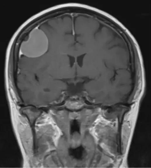
If your brain MRI report mentions a "dural tail," the phrase can sound alarming, but it is one of the more reassuring technical terms a radiologist can use. A dural tail is a pattern of contrast enhancement, not a diagnosis on its own, and it is best known as a helpful clue that a mass sitting just outside the brain tissue is very likely a meningioma, the most common primary brain tumor and one that is usually benign. This article covers what radiologists mean by a dural tail, what else can cause one, whether it points to something serious, and what typically happens next.
What does "dural tail" mean?
The dura mater is the tough outer membrane lining the inside of the skull and wrapping around the brain and spinal cord. When a mass sits against or grows from the dura, intravenous (gadolinium) contrast during an MRI can light up a thin line of enhancement trailing off the edge of the mass along the adjacent dura, tapering as it moves away, a bit like a comet's tail.
This pattern only shows up after contrast, so it lives on gadolinium-enhanced MRI. It can occasionally be seen on contrast CT, but MRI is far better at picking up thin, subtle enhancement.
- Appears as a thin, curved line of enhancement hugging the dura next to a mass, thickest close to the tumor and fading with distance
- Only visible after intravenous contrast, almost always on MRI
- Helps confirm a mass sits outside the brain tissue itself (an "extra-axial" location) rather than within it
What causes a dural tail sign?
By far the most common cause is a meningioma, a tumor that grows from the meninges, the layers of tissue covering the brain. Roughly two out of three meningiomas show a dural tail, which is why the sign is so closely linked with that diagnosis.
The tail can reflect two things: true tumor cells spreading a short distance along the dura, or a reactive response in nearby tissue, extra blood vessels and fibrous tissue without any tumor at all. Both patterns show up on pathology, so a longer tail does not necessarily mean a more extensive tumor.
Meningioma is far from the only cause, though. Other conditions that can produce the same look include:
- Metastatic tumors that have reached the dura from cancer elsewhere in the body
- Lymphoma involving the meninges
- Neurosarcoidosis and other inflammatory conditions that thicken the dura
- Solitary fibrous tumor (formerly hemangiopericytoma), a less common dural-based tumor
- Idiopathic hypertrophic pachymeningitis, a rare inflammatory thickening of the dura
- Reactive dural thickening after surgery, radiation, or a chronic subdural collection
Is a dural tail sign always serious?
No. A dural tail describes a contrast enhancement pattern, not how aggressive or dangerous a mass is. Even when a meningioma is the cause, the large majority are slow-growing and classified as benign (WHO grade 1), and plenty are small enough to simply be watched with periodic imaging rather than treated.
What matters more is everything else about the mass: its size, growth, location, and whether it causes symptoms. Your radiologist and your own physician weigh all of that together, not the tail in isolation.
What symptoms are linked to a dural tail sign?
Many masses that produce a dural tail are found by chance, on a scan ordered for something unrelated. Having no symptoms at all is common and does not mean anything was missed.
When symptoms do occur, they depend on where the mass sits and how large it has grown, not on the dural tail itself. Possibilities include headaches, seizures, changes in vision, weakness on one side of the body, or subtle personality or memory changes if the mass presses on the frontal lobes. Most people with a small, incidentally found mass never develop any of these.
How is a dural tail sign followed up?
Once a dural tail is noted, the focus shifts to the mass itself: its size, shape, location, and whether nearby bone or brain tissue shows any reaction.
For a small, asymptomatic mass with imaging features typical of a meningioma, the usual plan is a follow-up MRI around 6 to 12 months later, then longer intervals if it stays unchanged. Growth, new symptoms, or atypical features prompt a referral to neurosurgery, where options range from continued monitoring to surgery or targeted radiation.
Why a second read can help
A dural tail sits at the intersection of a memorable-sounding term and a genuinely important question: what exactly is this mass, and does anything need to be done about it right now? An expert radiologist reviewing your actual MRI images, not just the report, can confirm whether the mass and its dural tail fit the typical, reassuring meningioma pattern or whether the appearance deserves a closer look. DocOrbit provides an expert second read of your scan that you can share directly with your own doctor, so a term like dural tail gets interpreted alongside your full clinical picture. Our guide on when to get a second radiological opinion can help if you are weighing whether that is worth pursuing.
Does a dural tail always mean I have a meningioma?
No. A dural tail sign makes meningioma the leading possibility on the list, since around two-thirds of meningiomas show one, but other dural-based conditions can produce the same pattern, including metastases, lymphoma, and inflammatory conditions like neurosarcoidosis. Your radiologist weighs the tail alongside the mass's size, shape, and location before suggesting a diagnosis.
Is a dural tail sign dangerous?
Not on its own. It describes a pattern of contrast enhancement, not how aggressive or dangerous a mass is. Even when the underlying cause is a meningioma, the large majority are slow-growing and benign, and many are simply watched with periodic scans rather than treated.
Do I need surgery if my MRI report mentions a dural tail?
Usually not right away. The decision depends on the mass itself, not the tail: its size, location, whether it is growing, and whether it is causing symptoms. Small, incidental, meningioma-typical masses are commonly followed with repeat imaging, while larger or symptomatic ones are usually discussed with a neurosurgeon.
Can a dural tail sign show up on a CT scan?
Occasionally, but it is far more reliably seen on contrast-enhanced MRI, which has better soft-tissue contrast and can pick up thin enhancement along the dura that a CT scan can miss entirely.
Key takeaways
- A dural tail is a thin line of contrast enhancement trailing off a mass along the adjacent dura, seen on contrast-enhanced MRI
- It is most closely linked with meningioma, which shows this pattern around two-thirds of the time, but it is not exclusive to it
- The sign itself does not indicate danger; the size, growth, and behavior of the underlying mass matter far more
- Many masses that show a dural tail are found incidentally and cause no symptoms at all
- Follow-up usually means periodic MRI for small, typical masses, with neurosurgical input reserved for growth, symptoms, or atypical features
Your report probably contains more than this one finding. Our free radiology report explainer puts the whole thing into plain English and gives you a short list of questions to take to your doctor.
How do I have my MRI reviewed by another radiologist online?
Yes. Online radiology second opinions run entirely from home: you upload your images, with no CD to post and no second trip, and a subspecialist radiologist re-reads the study and writes a structured report you can give to your own doctor. DocOrbit returns that signed second read in 24 to 48 hours. You can then message the radiologist with questions about the MRI and about what the report actually means for you.
This article is for general information only and is not medical advice. Always discuss your imaging results and any next steps with a qualified physician.