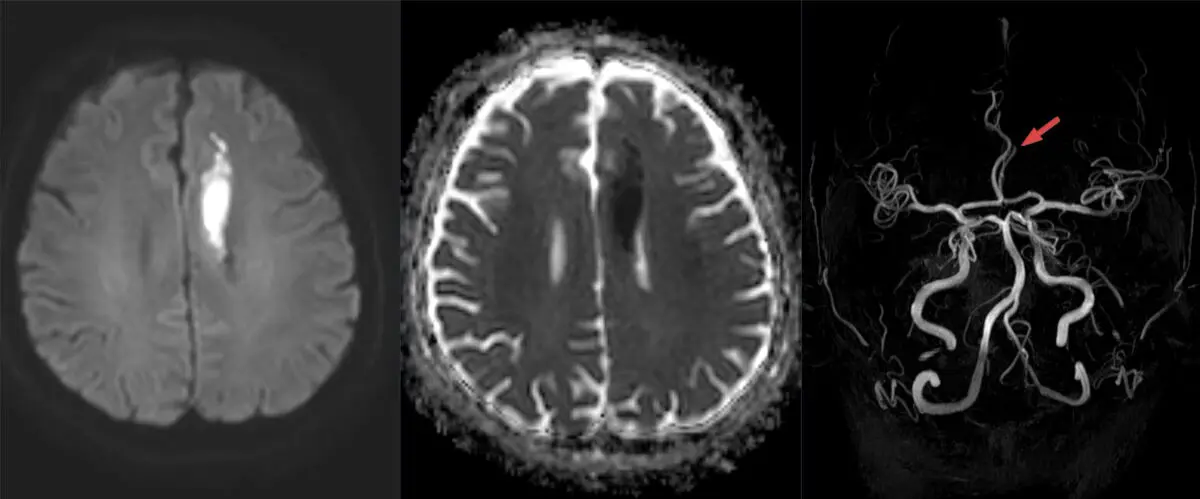
If your MRI report mentions "diffusion restriction" or "restricted diffusion," it helps to know what that phrase is actually describing: how freely water molecules are moving inside a small piece of tissue. It is a physics observation, not a diagnosis. The same finding can point to something urgent, something treatable, or nothing at all, depending on where it is and what brought you in for the scan. This article explains what radiologists are looking at, why they always check a second image before believing it, and what the finding usually means in context.
What does "diffusion restriction" mean?
Water molecules in living tissue are never still. They jiggle and drift in random directions, and how far they get in a fraction of a second depends on what is in their way. A diffusion-weighted sequence, usually abbreviated DWI, is an MRI setting tuned to measure exactly that movement.
Where water moves freely, such as in the fluid spaces of the brain, the signal fades. Where movement is hemmed in, the signal stays bright. That bright, hemmed-in appearance is what gets written up as diffusion restriction.
Broadly, water gets hemmed in for three reasons:
- Cells swell and squeeze out the space around them, which is what happens within minutes when blood flow to an area stops
- Cells are packed unusually tightly, leaving little room between them, as in some dense tumors and in normal lymphoid tissue
- The fluid itself is thick and sticky rather than watery, as with pus inside an abscess or older blood products
None of these three is inherently more likely than the others. Which one applies is a question the rest of the scan and your clinical picture answer, not the diffusion image alone.
Why the ADC map matters
A bright spot on DWI is not enough to call something restricted. Some tissue looks bright on the diffusion images simply because it is bright on the underlying images that the sequence is built on, an effect radiologists call T2 shine-through. It looks convincing and means nothing.
That is why every diffusion study comes with a companion image called the ADC map, which measures the same water movement numerically. Genuinely restricted tissue is bright on DWI and dark on ADC. If an area is bright on DWI but not dark on ADC, it is shine-through, and a careful report will say so.
This is also why reports often include a phrase like "restricted diffusion with corresponding low ADC values." It reads like jargon, but it is the radiologist explicitly confirming that the finding survived the check. Old scar tissue is a good example of the trap: areas of gliotic change from a healed injury can look bright on several sequences while diffusing water perfectly normally.
Common causes
Diffusion restriction appears in a wide range of situations. The most frequently reported include:
- Acute ischemic stroke, where restriction appears within minutes of the blood supply being interrupted and is the earliest reliable sign on MRI
- Abscess, in the brain or elsewhere, because pus is thick and restricts strongly
- Densely cellular tumors, including lymphoma, higher-grade brain tumors, and some metastases
- Epidermoid cysts, a benign lesion that characteristically restricts and is identified largely by this feature
- Active demyelinating plaques during their inflammatory phase
- Temporary changes after a prolonged seizure, after very low blood sugar, or in certain metabolic and toxic states
- Blood products from a recent bleed
- Artifact, particularly near the skull base, air spaces, dental work, or metal implants, where the sequence is prone to distortion
That last one is worth knowing about. Diffusion imaging is fast but relatively fragile, and an apparent restriction sitting exactly where the technique is known to misbehave is treated with healthy skepticism.
Is it serious?
Everything depends on the setting. In someone who arrived with sudden weakness, facial droop, or trouble speaking, a new area of restricted diffusion is essentially the imaging signature of an acute stroke, and it is handled as an emergency. In that context the finding is not incidental, and nobody waits for a follow-up scan.
In a routine outpatient MRI done for headaches or dizziness, the picture is usually quieter. A tiny dot of restriction may represent a very small vessel event, similar to what our explainer on a lacunar infarct on MRI describes, and the response is a workup of blood pressure, cholesterol, heart rhythm, and the neck vessels rather than emergency treatment.
Timing also carries information. Restriction from a stroke fades over roughly one to two weeks as the tissue evolves, so its presence suggests the event was recent and its absence suggests an older one. Diffusion restriction itself causes no symptoms; any symptoms come from whatever is producing it, and plenty of people with a small restricting area notice nothing at all.
Diffusion restriction outside the brain
Diffusion imaging started in neuroradiology but is now routine in the abdomen, pelvis, breast, and whole-body MRI. In the prostate it is one of the sequences behind PI-RADS scoring; in the liver and lymph nodes it helps flag areas worth a second look; in the pelvis it can help separate an abscess from a simple fluid collection.
Outside the brain, though, the finding is much less specific. Normal spleen, tonsil and lymphoid tissue, bowel, testes, and endometrium all restrict diffusion perfectly normally. Here it works more like a "look closer" marker than a verdict, in much the same way a hypermetabolic focus on a PET-CT flags activity without naming its cause. It is read alongside contrast images and the other sequences, never on its own.
How it is worked up
What happens next follows from the suspected cause rather than the word itself. When a stroke is likely, the next steps are usually imaging of the neck and brain vessels, heart rhythm monitoring, and blood tests looking for the reason. When an abscess is a possibility, contrast images and blood work usually follow, sometimes with additional MRI techniques that can help separate pus from tumor.
And when the finding is subtle, ambiguous, or sits in a spot where artifact is common, the most useful next step is often simply a repeat MRI after a short interval. Follow-up intervals vary by patient and by suspected cause, and your own doctor is the one who knows your history well enough to set them.
Why a second read can help
Diffusion restriction is one of the findings where interpretation genuinely changes the answer. Whether the ADC map was checked, whether the area sits in a known artifact zone, and whether it matches your symptoms all determine what the finding means, and those judgments come from the reader, not the machine. DocOrbit offers an expert second read of your scan that you can share directly with your care team, which can be worth having when a report leaves you unsure how much weight a single line deserves. If you are weighing that up, our guide on when to get a second radiological opinion is a good starting point, along with our broader look at the role of second opinions in radiology.
Does diffusion restriction always mean a stroke?
No. A stroke is the most familiar cause and the most time-sensitive one, which is why it is considered first when the symptoms fit. But abscesses, some tumors, active inflammation, seizures, and even scanning artifacts can all restrict diffusion. Your symptoms, their timing, and the rest of the MRI decide which explanation fits.
What is the difference between DWI and the ADC map?
DWI is the raw diffusion image, where restricted areas look bright. The ADC map is a calculated image that measures the same water movement numerically, and truly restricted tissue looks dark on it. Radiologists check both because an area can look bright on DWI simply because it is bright on the underlying images, an effect called T2 shine-through, which is not real restriction.
Does diffusion restriction mean cancer?
Not on its own. Densely packed tissue restricts diffusion, and some tumors are densely packed, but so are normal lymph nodes, tonsil tissue, the spleen, and the pus inside an ordinary abscess. Diffusion is one clue among several, and it is always read alongside contrast behavior, shape, location, and the other sequences before anything is concluded.
Can diffusion restriction go away?
Yes, often. Restriction after a stroke typically fades over one to two weeks as the tissue changes, a process radiologists call pseudonormalization, and restriction caused by a seizure, low blood sugar, or short-lived inflammation can disappear completely. That is one reason a repeat MRI is sometimes more informative than the first one.
What does diffusion restriction mean on a prostate or abdominal MRI?
Outside the brain, diffusion imaging is used as a supporting sequence rather than a standalone answer. It helps flag areas worth a closer look in the prostate, liver, lymph nodes, and pelvis, but plenty of normal tissue restricts too. In these regions the finding is combined with contrast images and scoring systems such as PI-RADS before it carries any weight.
Key takeaways
- Diffusion restriction describes water molecules moving less freely than expected in a piece of tissue. It is a measurement, not a diagnosis
- It is only believed when the bright DWI area is also dark on the ADC map, which rules out the common look-alike called T2 shine-through
- Acute stroke is the most urgent cause, but abscess, dense tumors, inflammation, seizures, and artifact all produce the same appearance
- Restriction from a stroke fades over one to two weeks, so its presence or absence helps date the event
- Outside the brain it is a supporting clue rather than an answer, since normal lymphoid tissue, spleen, and bowel restrict too
This article is for general information only and is not medical advice. Always discuss your imaging results and any next steps with a qualified physician.