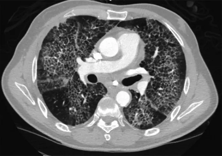
Seeing the phrase "crazy paving" in a chest CT report is jarring, mostly because it sounds like it belongs in a landscaping catalog rather than a medical document. Here is the useful starting point: crazy paving is a description of a pattern the radiologist can see, not a diagnosis, and the list of things that produce it is dominated by conditions doctors treat routinely. This article explains what radiologists mean by the term, what causes it, how much weight it carries, and what usually happens next.
What does "crazy paving" mean?
The pattern is made of two ingredients layered on top of each other. The first is a hazy, veil-like grayness over part of the lung, the finding radiologists call ground-glass opacity. The second is a network of thin white lines drawn across that haze, which are the normally invisible partitions between the smallest units of lung tissue, thickened enough to become visible.
Put together, the haze and the lines look like irregular paving stones set into pale mortar, which is where the name comes from. It is a thin-section CT finding and essentially never something you would see on a plain chest X-ray, because the structures involved are only a millimeter or two across.
What the pattern tells the radiologist is that both compartments of the lung have something in them at the same time: the air sacs themselves, and the scaffolding between them. That "something" can be fluid, blood, protein-rich material, inflammatory cells, or infection, which is precisely why the term on its own does not name a disease.
Several details around the pattern carry far more meaning than the phrase itself, and a good report usually notes them:
- How much of the lung is involved, and whether it is one patch or spread through both lungs
- Where it sits, since fluid from the heart tends to favor the lower and central lung while some other causes favor the upper zones
- Whether the affected areas have sharp geographic borders against normal lung, or fade gradually
- What else is on the scan, such as fluid around the lungs, an enlarged heart, or enlarged lymph nodes
- How it compares with any earlier scans, and how quickly it appeared
Common causes
Crazy paving has an unusually broad list of causes for a single pattern, which is one reason radiologists are careful not to over-interpret it. Common explanations include:
- Fluid building up in the lungs, most often because the heart is not pumping efficiently, and sometimes from other causes of leaky lung capillaries
- Viral pneumonia, including COVID-19, which made this pattern familiar to a whole generation of clinicians
- Pneumocystis pneumonia, an infection seen mainly in people whose immune system is suppressed
- Some bacterial pneumonias, particularly when they are extensive
- Bleeding into the air sacs, which can follow certain inflammatory conditions or blood-thinning medication
- Acute lung injury of any kind, including the severe form doctors call ARDS
- Organizing pneumonia, an inflammatory healing response that can follow an infection, a medication, or radiation treatment
- Lipoid pneumonia, when oily substances reach the lung, classically from long-term use of oil-based nose drops or laxatives
- Pulmonary alveolar proteinosis, the textbook cause named in every radiology lecture, in which protein-rich material accumulates in the air sacs. It is genuinely rare, and it is not the most likely answer simply because it is the most quoted one
- Uncommonly, a slow-growing lung cancer that spreads along the surfaces of the air spaces rather than forming a lump
Because the same appearance covers everything from a treatable chest infection to a rare metabolic lung condition, timing and symptoms do most of the diagnostic work. If your report also describes hazy areas without the overlying lines, our explainer on what ground-glass opacity means covers the underlying finding in more detail.
Is it serious?
An honest answer: crazy paving usually reflects something active in the lungs rather than an old scar, so it is a finding doctors follow up on rather than file away. In practice it most often turns up in people who are already unwell and already being investigated, so the scan tends to confirm that something is going on rather than reveal a surprise.
Active is not the same as dangerous. Fluid from the heart and ordinary pneumonia are both very treatable, and in both the pattern typically clears once the cause is dealt with. Doctors look harder when oxygen levels are low, when the pattern spreads quickly over days, when the immune system is suppressed, or when it is still there months later without an explanation. As always, what the pattern means depends on your symptoms, exam, and history rather than on the images alone.
Symptoms
The pattern itself causes nothing. Any symptoms come from whatever is filling the lung. Depending on the cause, that might mean:
- Breathlessness, especially on exertion, which is the most common accompaniment
- A cough, dry or productive
- Fever, chills, or feeling generally unwell when an infection is behind it
- Reduced oxygen levels, sometimes noticed first on a finger probe rather than felt
- Slow-building fatigue and breathlessness over months, in the rarer chronic causes
- Occasionally nothing at all, when a limited area turns up on a scan ordered for something else
How is it diagnosed and followed up?
The single most useful clue is not on the scan at all: it is how long this has been going on. A pattern that appeared over two days points in a very different direction than one that has built up over six months, and doctors use that split to decide what to test first.
From there the workup follows the suspected cause. Blood tests and oxygen measurement are near-universal. A heart assessment such as an echocardiogram is added when fluid overload is on the list, and swabs or sputum samples when infection is. Bronchoscopy with a gentle lung washing is reserved for the cases that stay unclear, though it is the test that clinches a diagnosis of alveolar proteinosis, because the washing fluid has a characteristic milky appearance. A repeat CT after treatment is the most common step of all, since a pattern that has cleared usually needs nothing further. The same unhurried, context-first approach applies to other chest findings, and our guides to the tree-in-bud pattern and to fluid around the lung show how a descriptive term gets read alongside the rest of the picture.
Why a second read can help
Crazy paving is a good example of a finding where the distribution, the pace, and the company it keeps narrow a long differential down to a short one, and where a careful reader adds real value. An expert read can help separate the pattern that fits straightforward heart failure or a resolving pneumonia from the one that deserves a bronchoscopy and a specialist opinion, and it gives you something concrete to bring to your own doctor. DocOrbit offers an expert second read of your scan that you can share directly with your care team, so the finding is interpreted in the context of your full history. If you are weighing whether to seek one, our guide on when to get a second radiological opinion may help, and you can read more broadly about the role of second opinions in radiology.
Does crazy paving on a CT scan mean I have COVID-19?
Not by itself. COVID-19 became one of the most familiar causes of the pattern, and it is often mentioned for that reason, but heart-related fluid in the lungs, other viral and bacterial infections, and several non-infectious conditions produce exactly the same appearance. A swab or blood test settles the question far more reliably than the scan does.
Is a crazy-paving pattern a sign of lung cancer?
Rarely. The pattern reflects material filling the air sacs and the tissue between them, which is the signature of fluid, infection, or inflammation rather than of a tumor. One uncommon slow-growing lung cancer spreads along the air spaces and can imitate it, which is why a pattern that persists for months without an explanation gets looked into. For most people the cause is something far more ordinary and often temporary.
Can a crazy-paving pattern go away?
Yes, and in many people it does. When the cause is fluid from the heart or an infection, the pattern often clears within days to weeks once the underlying problem is treated. That is exactly why a repeat CT after treatment is such a common next step, since a scan that has cleared usually closes the question.
How serious is a crazy-paving pattern on a CT scan?
It depends entirely on the cause, how much of the lung is involved, and how you feel. The pattern usually reflects something active in the lungs rather than an old scar, so it is generally taken seriously and investigated promptly. Active is not the same as dangerous, though, and the most common causes are treatable conditions that resolve.
What tests are done after crazy paving is found on a CT scan?
The workup follows the suspected cause rather than the pattern. Blood tests, oxygen measurement, swabs or sputum samples, and a heart assessment such as an echocardiogram cover most situations. When the picture stays unclear, or when a rarer cause is suspected, doctors may add a bronchoscopy with a lung washing, and they will usually repeat the CT once treatment has had time to work.
Key takeaways
- Crazy paving describes hazy lung crossed by a network of thickened lines on a CT scan. It is a description of an appearance, not a diagnosis
- The list of causes is broad and includes fluid from the heart, pneumonia, bleeding, and inflammation. The famous rare cause, alveolar proteinosis, is rarely the answer
- How fast the pattern appeared matters more than the pattern itself, and it is what steers the first round of tests
- Most common causes are treatable, and the pattern often clears entirely on a repeat scan after treatment
- Low oxygen, rapid spread, a suppressed immune system, or a pattern that persists for months all prompt a closer look
Your report probably contains more than this one finding. Our free radiology report explainer puts the whole thing into plain English and gives you a short list of questions to take to your doctor.
Can I ask a radiologist about my CT report online?
Yes. Online radiology second opinions run entirely from home: you upload your images, with no CD to post and no second trip, and a subspecialist radiologist re-reads the study and writes a structured report you can give to your own doctor. DocOrbit returns that signed second read in 24 to 48 hours. You can then message the radiologist with questions about the CT and about what the report actually means for you.
This article is for general information only and is not medical advice. Always discuss your imaging results and any next steps with a qualified physician.