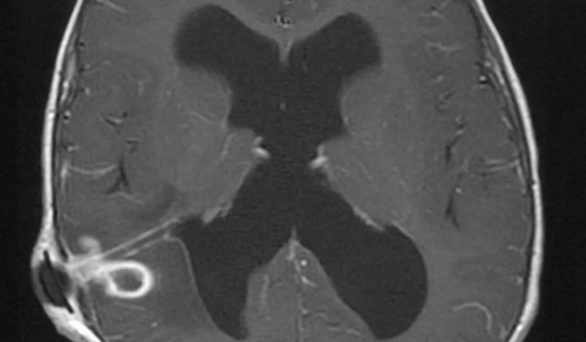
If your MRI or CT report says that something "enhances" or shows "contrast enhancement," it is describing one specific thing: that area got brighter after a contrast agent was injected into a vein. That is a statement about blood supply, not a diagnosis. Plenty of entirely normal structures enhance on every scan, and some findings that do need attention barely enhance at all. This article explains what radiologists are actually seeing, what the different enhancement patterns tend to suggest, and why the word on its own does not tell you whether a finding is worrying.
What does "contrast enhancement" mean?
Contrast agents are liquids that change how tissue looks on a scan. They are not dyes in the everyday sense and they do not stain anything permanently. They circulate in the blood, spread into tissue for a while, and are then cleared, mostly by the kidneys.
Which agent is used depends on the machine:
- CT uses an iodine-based agent, which absorbs X-rays strongly and makes well-supplied tissue look denser and whiter
- MRI uses a gadolinium-based agent, which changes the magnetic behavior of nearby water and makes well-supplied tissue look brighter on T1-weighted images
- Ultrasound can use tiny gas microbubbles, which reflect sound waves and light up flowing blood in real time
Enhancement simply means that a region took up more of that agent than the tissue around it. Two things drive it: how much blood is being delivered to the area, and how leaky the small vessels there are. Tissue with a rich blood supply and leaky capillaries enhances strongly. Tissue with almost no blood supply, such as the fluid inside a simple cyst, does not enhance at all.
Why some tissue enhances and some does not
In most of the body, small blood vessels are naturally somewhat leaky, so contrast seeps into the surrounding tissue to some degree everywhere. The brain is the important exception. Its vessels are sealed by the blood-brain barrier, so contrast normally stays inside them.
That is why enhancement inside brain tissue carries extra meaning: it implies that the barrier has been disturbed by a tumor, inflammation, infection, or a recent injury. Even here, some structures enhance normally because they sit outside the barrier, including the pituitary gland, the choroid plexus inside the ventricles, the dura covering the brain, and the lining of the nose and sinuses. Seeing those light up is expected, not a finding.
Radiologists therefore rarely describe enhancement as simply present or absent. They describe how much, where, in what shape, and at what moment after injection.
The patterns radiologists describe
Most of the interpretive weight sits in the pattern rather than the brightness. The ones that appear most often in reports include:
- Homogeneous enhancement — the whole lesion brightens evenly, typical of well-organized, well-supplied tissue such as a meningioma or a nerve sheath tumor
- Heterogeneous enhancement — patchy, uneven brightening, often because parts of the lesion have outgrown their own blood supply
- Ring or rim enhancement — a bright edge around a dark center, seen with abscesses, some metastases, higher-grade tumors, and active demyelinating plaques
- Peripheral nodular enhancement with fill-in — bright dots at the edge on early images that gradually fill the lesion on later ones, the near-signature pattern of a liver hemangioma
- Avid or early arterial enhancement — the lesion brightens fast, before the surrounding organ, indicating a direct arterial supply
- Delayed or persistent enhancement — the lesion stays bright late, which is more typical of fibrous or scar-rich tissue
- No enhancement — the area looks the same before and after, typical of simple cysts and plain fluid
Reports often also mention a dural tail, a thin streak of enhancement extending from a mass along the covering of the brain, or washout, where a lesion brightens early and then becomes darker than the organ around it. Both are shorthand for a behavior over time rather than a single snapshot.
Does timing change the answer?
Frequently, yes. Many CT and MRI protocols acquire several sets of images after the injection: an arterial phase within seconds, a venous or portal phase after roughly a minute, and sometimes a delayed phase several minutes later.
A finding can look completely different at each of those moments, and it is the sequence of behaviors that carries the diagnostic information. This is why a liver or pancreas study is sometimes repeated with a specific multi-phase protocol: not because the first scan was faulty, but because the earlier study caught the finding at only one point in time.
Does an enhancing finding mean something serious?
Not by itself. Enhancement is one of several features a radiologist weighs, alongside size, borders, internal texture, how the finding behaves on other sequences such as diffusion-weighted imaging, and your clinical history. A brightly enhancing area in someone with a fever and rising inflammatory markers usually reads very differently from the same appearance in a healthy person having a scan for an unrelated reason.
Several genuinely benign situations enhance strongly: infection and abscess, active inflammation, healing tissue after surgery or injury, normal glands, and common non-cancerous growths such as fibroids and hemangiomas. Meanwhile some slow-growing tumors enhance only faintly. Using enhancement alone as a proxy for "serious or not" gives the wrong answer in both directions.
What about the contrast agent itself?
Contrast agents in routine use today have a good safety record, and the department will normally check a few things before the injection: kidney function, previous reactions to contrast, asthma or significant allergies, and whether you are pregnant or breastfeeding. Mild, short-lived sensations such as warmth spreading through the body or a metallic taste are common and expected.
Gadolinium is retained in very small amounts in some tissues after repeated MRI studies. No health effect has been established from this, but it is one reason contrast is used when it answers a question and skipped when it does not. If your report notes that a study was performed without contrast, that describes the chosen technique rather than a shortcoming, though a follow-up study with contrast is sometimes recommended when a finding needs further characterization.
Why a second read can help
Enhancement is one of those findings where the interpretation, rather than the image, carries most of the weight. Whether the pattern is truly nodular or merely patchy, whether the timing was right for the question being asked, and whether an area is enhancing at all or just looked bright beforehand are all judgment calls made by the reader. DocOrbit offers an expert second read of your scan that you can share directly with your own doctor, which many people find useful when a single line in a report changes the plan. If you are weighing that up, our guide on when to get a second radiological opinion and our wider look at the role of second opinions in radiology are good places to start.
Does contrast enhancement mean cancer?
No. Enhancement only means an area received contrast through its blood supply, and inflammation, infection, healing tissue, scar, and many harmless growths all do that. Normal structures such as the pituitary gland, the lining of the nose, blood vessels, and the covering of the brain enhance on every study. The pattern, shape, location, and timing decide what the enhancement suggests, not the fact that it happened.
What does no contrast enhancement mean?
It usually means the area has no meaningful blood supply of its own, which is reassuring in many settings. Simple cysts, old scar tissue, and plain fluid collections typically show no enhancement at all, and that is one of the features used to call them benign. It is not a guarantee on its own, since a few slow-growing or low-blood-flow lesions enhance very little, so it is read together with size, shape, and the other sequences.
What is a ring-enhancing lesion?
It is an area whose rim lights up after contrast while the center stays dark, which happens when the outer edge has an active blood supply and the middle does not. The classic causes are an abscess, a metastasis, a higher-grade brain tumor, and an active demyelinating plaque, and these can often be separated by the thickness and regularity of the ring plus the other sequences. It is a finding doctors look into promptly, but it is not a diagnosis by itself.
Is the contrast dye used for MRI and CT safe?
For most people it is well tolerated, and the agents used today have a good safety record. CT uses an iodine-based agent and MRI uses a gadolinium-based one, and both are usually checked against your kidney function, allergy history, and pregnancy status before injection. Mild reactions such as warmth or a metallic taste are common and brief, and serious reactions are uncommon and the department is equipped for them.
Why do some scans need contrast and others do not?
Contrast is added when the question being asked depends on blood supply, such as characterizing a mass, looking for infection or inflammation, or mapping vessels. For many other questions, including kidney stones, most fractures, and routine lung imaging, contrast adds nothing and is deliberately left out. A report that says the study was done without contrast is describing the technique used, not something that was missed.
Key takeaways
- Contrast enhancement means an area got brighter after an injection, which reflects blood supply and vessel leakiness rather than any single diagnosis
- Normal structures including the pituitary gland, dura, nasal lining, and blood vessels enhance on every scan
- The pattern matters far more than the brightness: even, patchy, ring-shaped, or filling in from the edge all point in different directions
- Timing is part of the finding, which is why multi-phase CT and MRI protocols exist for the liver, pancreas, and kidneys
- Absent enhancement is often reassuring, but it is interpreted alongside size, borders, and the rest of the study rather than on its own
This article is for general information only and is not medical advice. Always discuss your imaging results and any next steps with a qualified physician.