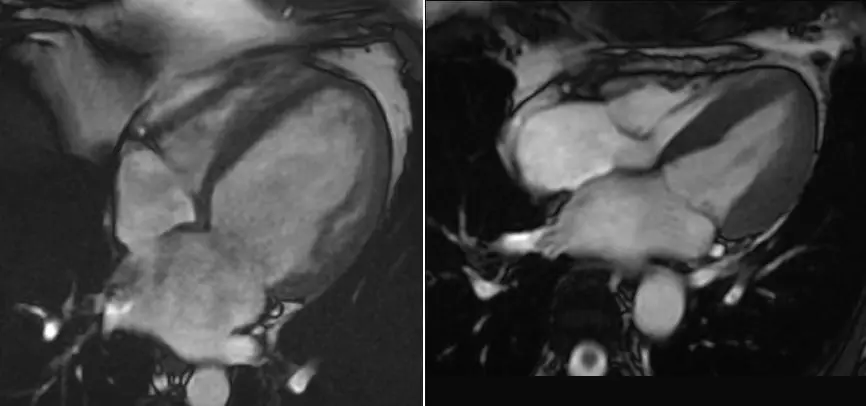
You already have the scan. Maybe a cardiologist ordered it after palpitations, an unclear echocardiogram or a family history of heart muscle disease, or after a heart attack to see how much muscle survived. Maybe the report uses terms like "late gadolinium enhancement" or "ejection fraction" that nobody has fully explained, or it came back normal and your symptoms haven't. A cardiac MRI second opinion, also called a heart MRI second opinion, means a board-certified radiologist reviews your existing images independently and writes a report you can bring to your cardiologist. Here is what that review covers and why this scan rewards a slower look.
A cardiac MRI holds far more than one set of pictures
The heart never stops moving, so the scanner is synchronized to your heartbeat and the study is built from many sequences. Most are short movies, called cine images, of the heart beating, repeated slice by slice and in several views. Others show the tissue itself: swelling, scar after contrast dye, and sometimes blood flow through a valve or into the muscle under stress. A full study often runs to well over a thousand frames. Numbers like ejection fraction (the share of blood the heart pumps out per beat) come from tracing the heart muscle on every slice, so two careful readers can differ slightly.
In a busy department the cardiac MRI interpretation is usually written around the question on the request form, such as "is there scar?" or "any sign of myocarditis?", and it answers that well. It is less suited to a small finding nobody was asking about: a thin line of enhancement in an unexpected part of the wall, mild pericardial thickening, or something at the edge of the picture such as a lung base or a lymph node. None of this reflects badly on whoever read it under time pressure; it is what volume and time do to any read of a study this dense. A slower second look buys that attention back, as our article on whether radiologists can miss things on MRI or CT scans explains.
Why a cardiac imaging specialist reads a heart MRI differently
A general radiologist reads everything in a single shift: chests, abdomens, spines, brains. Cardiac imaging is its own subspecialty, a corner of cardiothoracic radiology, and in many hospitals heart MRI is read by cardiologists or radiologists trained specifically in it. Where scar sits in the heart wall is a diagnostic clue: scar that starts on the inner surface and spreads outward within one artery's territory points toward a blocked coronary artery, while a stripe through the middle of the wall or along the outer surface points toward other causes, such as myocarditis or certain cardiomyopathies (diseases of the heart muscle).
Experience also protects against over-calling. Prominent muscle ridges in a healthy or athletic heart can be mistaken for noncompaction, fluid in a normal pericardial recess can pass for a lymph node, and motion, an irregular rhythm or a nearby device can create artifacts that mimic scar. That is the logic behind asking a subspecialist for a cardiac MRI second read: not that the first reader was careless, but that a narrower, deeper focus catches patterns a broad one is more likely to under-call, and recognizes harmless variants for what they are.
What a cardiac MRI actually shows, structure by structure
A thorough cardiac MRI review works through the same anatomy a cardiac imaging specialist checks on every study:
- The left ventricle: size, wall thickness, ejection fraction and wall motion; thickened walls (hypertrophic cardiomyopathy, long-standing high blood pressure, athlete's heart), a stretched, weakened ventricle (dilated cardiomyopathy), and the footprint of coronary disease, including atherosclerotic plaque in the arteries that feed it
- The heart muscle tissue: after contrast, scar (late gadolinium enhancement) and swelling are mapped by pattern and location, separating a past heart attack from myocarditis, sarcoidosis or amyloid; the less of the wall thickness is scarred after a heart attack, the likelier that muscle is to recover if blood flow is restored
- The right ventricle and atria: chamber size, pumping function and wall motion, strain from lung disease, masses such as a myxoma and, less often, arrhythmogenic cardiomyopathy
- The valves: cine and flow measurements estimate how much blood leaks backward (regurgitation) or how tight a valve is (stenosis); echocardiography is usually first, and MRI earns its place when the echo is borderline
- The pericardium: thickening, fluid (pericardial effusion), inflammation, and signs of constriction, where the sac around the heart stiffens
- Masses and clots: how tissue behaves on different sequences helps separate fat, fluid, clot and tumor; a clot inside the left ventricle after a heart attack is a classic MRI catch, a dark patch with no blood supply against bright blood
- The aorta and great vessels: the aortic root, ascending aorta and pulmonary artery are measured, and coarctation (a narrowing of the aorta) and congenital shunts assessed; the whole aorta needs a dedicated MR angiogram
- Everything else in view: the lung bases, a pleural effusion, lymph nodes, chest wall and upper abdomen sit at the edges of the images and are easiest to overlook
Three limits matter. MRI shows what coronary disease does to the heart muscle, but not reliably the artery narrowing itself or calcium in its wall; CT is the usual test for that, as our coronary artery calcium score article explains, while a stress perfusion MRI asks the same question as a myocardial perfusion scan by a different technique. A heart MRI also images the lungs poorly, so it cannot clear a lung nodule; that takes a chest CT, covered in our thorax CT second opinion guide. And a second read is only as good as the images: if a rhythm problem or poor breath-holds spoiled a sequence, a careful reader says so rather than guessing.
How DocOrbit reads your cardiac MRI
Upload your scan for a second opinion, the DICOM files from the study or a copy of the disc, and a board-certified radiologist matched to your scan performs a full cardiac MRI second read, going through the same structures one at a time and writing a structured report you can hand directly to your cardiologist. Include every series if you can, cine loops and contrast images as well as key pictures, because a heart MRI cannot be judged from stills. The signed report comes back in 24–48 hours. It is not a verdict on the first read, only what a fresh, unhurried look at your heart MRI found. You can have your cardiac MRI re-read by another radiologist starting today.
Get your cardiac MRI re-read by a board-certified radiologist
Ask the radiologist questions afterward
The second opinion does not end when the report lands. Once you have it, you can ask a radiologist online directly, about a phrase in the report, whether a finding explains your symptoms, or what to raise with your cardiologist next, and get a written answer.
Or get an answer in minutes with Orbius
If waiting 24–48 hours doesn't suit you, DocOrbit also offers Orbius, an AI radiology agent built for exactly this kind of online radiology second opinion. It reads the same cardiac MRI and returns a structured report of findings, conclusion and next steps in minutes, at a lower cost, at any hour. It is an honest trade of speed and price against a specialist's signature. The two are separate options, not two steps of one service: if you later want a radiologist, you can book one with a click and your images already loaded, but that is a separate consultation in which the radiologist reads the scan from scratch and writes their own report, without reviewing or signing Orbius's.
How do I send my cardiac MRI for a second opinion?
Upload the DICOM files from your scan, or the CD you were given at the imaging center or hospital, directly through the DocOrbit platform, and include every series, cine loops and contrast images too. Nothing needs to be mailed, and the images go straight to the radiologist reading your case.
Will my cardiologist accept a second opinion report?
Usually, yes. The report follows the structured format radiologists use everywhere, with findings and an impression your cardiologist can read like any other imaging report, and you are free to share it. Your cardiologist knows your history and puts the report in context.
How long does a cardiac MRI second opinion take?
A signed report from a board-certified radiologist typically comes back in 24 to 48 hours. If you want an answer sooner, Orbius, DocOrbit's AI radiology agent, returns a structured report on the same images in minutes.
Is it worth getting a second opinion if my cardiac MRI was reported as normal?
It can be, especially if your symptoms haven't settled or the report mentions a borderline finding without a clear next step. A fresh, unhurried review sometimes turns up something the first read wasn't looking for, and just as often confirms that normal really does mean normal.
Key takeaways
- A cardiac MRI is many sequences, mostly beating-heart movies and contrast images, often read at speed for one question
- Cardiac imaging is a subspecialty, and experience shows in telling scar patterns, normal variants and artifacts apart
- A proper second read covers each structure and says what MRI cannot show, such as coronary calcium or the lungs
- DocOrbit's signed radiologist read takes 24–48 hours; Orbius, the AI option, returns a structured report in minutes
- You can message the radiologist with questions after the report arrives
Can I get a second opinion on my cardiac MRI online?
Yes. You upload your images from home, with no CD to mail and no second trip to the imaging center, and a board-certified radiologist matched to your scan re-reads the study and writes a structured report you can hand to your own doctor within 24 to 48 hours. You can message the radiologist with questions afterward, and Orbius can return a first read in minutes if you want an answer sooner.
This article is for general information only and is not medical advice. Always discuss your imaging results and any next steps with a qualified physician.
