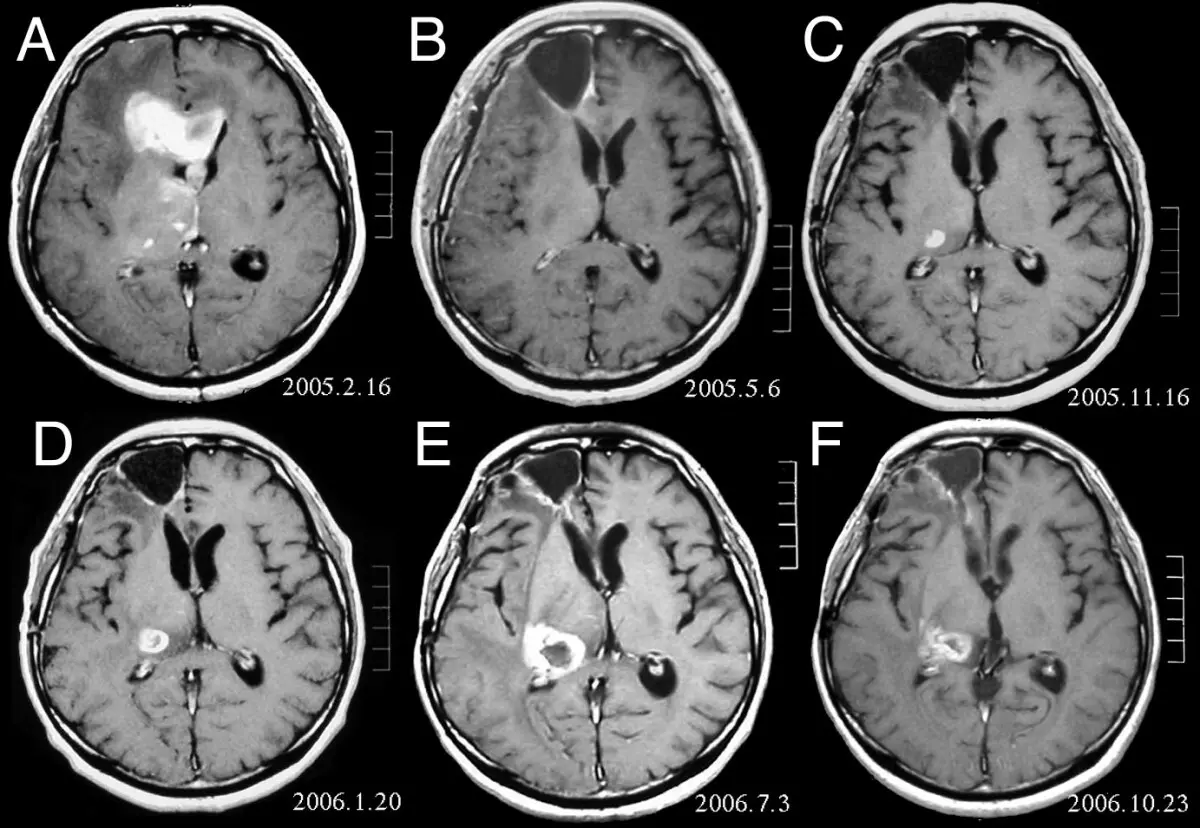
Yes, MRI (magnetic resonance imaging, a scan using a strong magnet and radio waves to build images) can detect many kinds of cancer, and for some organs it's one of the sharpest tools radiology has. But it can't do everything: no single scan, MRI included, finds every cancer, and none can diagnose one on its own. An MRI can show a mass is there and describe how it looks, but only a biopsy (a small tissue sample examined under a microscope) confirms what it actually is. Here's what MRI is genuinely good at, where it isn't the first choice, and what usually happens next.
What MRI is good at finding
MRI's strength is soft-tissue contrast, its ability to tell subtly different soft tissues apart on the same image, better than CT or an X-ray can. That makes it especially valuable for cancers in and around:
- Brain and spinal cord
- Breast tissue, particularly when it's dense enough that mammograms are less reliable
- The prostate, where multiparametric MRI (several MRI sequences combined for a sharper look) often precedes a biopsy
- The liver, where MRI helps tell a harmless hemangioma (a benign tangle of blood vessels) from something more concerning
- Soft tissue and bone marrow, including sarcomas (cancers of fat, muscle, or connective tissue)
Contrast dye (gadolinium, injected into a vein) makes many of these tumors easier to spot, since cancers build their own, leakier blood supply, so more dye pools inside them than the surrounding tissue.
Where MRI isn't the best first choice
The lungs are mostly air, which gives no useful MRI signal, so CT is far more sensitive for lung nodules and lung cancer; our guide on what to do after a lung nodule shows up on CT covers that in detail. Once cancer has spread widely through bone, a bone scan (a nuclear medicine test) or a PET-CT usually images the whole skeleton faster than MRI can, slice by slice. When hunting the whole body for a cancer of unknown origin, CT or PET-CT is typically the more practical start.
None of this makes MRI a lesser tool, it just means the "best" scan depends on the organ and the question, which is why oncology teams routinely combine imaging types.
Why no scan alone can diagnose cancer
What MRI actually measures is a lesion's shape, size, how smooth or irregular its edges are, and how it responds to contrast, patterns that make a finding look "probably benign," "suspicious," or "indeterminate." None of those words is a diagnosis. Some benign conditions mimic cancer closely on imaging, and some early cancers look unremarkable, which is why radiologists deliberately hedge rather than call it either way from pictures alone.
Think of an MRI like a metal detector on a beach: it can tell you something is buried and roughly how big it is, but not whether it's a gold coin or a bottle cap. Working that out takes digging it up, in medicine that's usually a biopsy, or a repeat scan months later to see if it has changed.
What happens after MRI finds something suspicious
The next step depends on what was found and where. Small, low-risk-looking findings are often just watched with a follow-up scan in a few months rather than biopsied right away, plenty of incidental findings turn out to be nothing. A more concerning one typically leads to a biopsy, sometimes a staging scan like PET-CT, and a referral to a specialist, often with a tumor board weighing in before treatment decisions are made.
If your report leans on hedging language, "cannot exclude malignancy" is a common one, it's worth having a subspecialty radiologist look at the actual images again rather than just the wording. That's the independent second opinion DocOrbit provides within a day or two, also the focus of our piece on how second opinions improve cancer outcomes.
Can MRI tell the difference between a benign and a malignant tumor?
Often, yes, to a useful degree. Radiologists weigh a lesion's shape, edges, and contrast pattern to judge how likely it is to be cancer, and some findings, like a simple cyst, are reliably benign on imaging alone. Many others fall into a gray zone only a biopsy can resolve.
Does a normal MRI mean I don't have cancer?
It's reassuring, but not an absolute guarantee. A normal MRI means no suspicious finding was seen in the scanned area with that technique. Very small or early lesions can be missed, and some cancers show up better on a different scan. Persisting symptoms may still warrant follow-up testing.
Is MRI with contrast better at finding cancer than MRI without contrast?
For most tumor detection, yes. Contrast dye highlights the abnormal, leaky blood vessels that many cancers grow to feed themselves, which makes some lesions far easier to see and characterize. Not every MRI needs contrast, though; your radiologist decides based on what's actually being looked for.
Why would a doctor order a CT or PET scan instead of an MRI?
Each scan is built for a different job. CT is faster and reads lungs, bone, and acute bleeding better than MRI does; PET-CT shows how metabolically active tissue is across the whole body at once, useful for staging. Doctors pick whichever tool answers the specific question in front of them.
Key takeaways
- MRI detects many cancers well, especially in the brain, spine, breast, prostate, and liver
- For lungs and widespread bone disease, CT, PET-CT, or a bone scan usually see more
- No scan, MRI included, diagnoses cancer by itself; a biopsy is typically needed
- A normal MRI is reassuring but doesn't rule out cancer with total certainty
- Words like "suspicious" or "indeterminate" describe probability, not a diagnosis
This article is for general information only and is not medical advice. Always discuss your imaging results and any next steps with a qualified physician.