
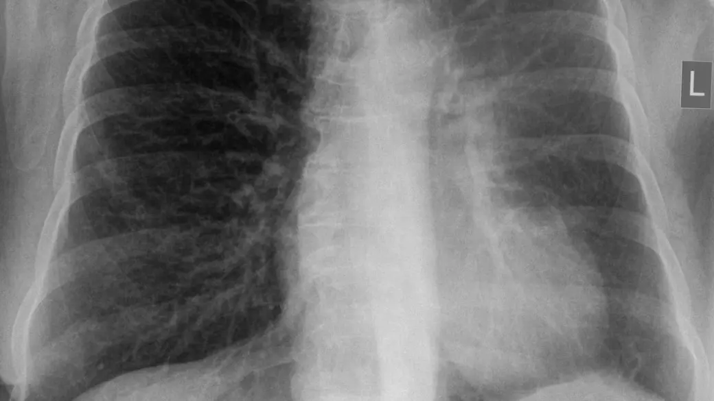
Reading the word "atelectasis" on a chest X-ray or CT report can sound alarming, especially because it is often described as a "collapsed" part of the lung. The reassuring truth is that atelectasis is very common, frequently small, and in most people clears up once its cause is dealt with. It describes how part of the lung looks on the scan, not a diagnosis on its own. Here is what it means, what causes it and how it is followed up.
What does "atelectasis" mean?
Your lungs are made of millions of tiny air sacs, called alveoli, that fill with air every time you breathe in. Atelectasis means some of them have deflated, so a section of lung holds less air than it should. The tissue is still there and still alive; it is simply not fully inflated. It can involve a small patch, a whole lobe or, less often, an entire lung.
It is not the same as a pneumothorax, where air has leaked into the space around the lung, although both are often called a "collapsed lung".
On imaging, radiologists look for a few things:
- On a chest X-ray, the deflated area looks whiter (denser) than the air-filled lung around it, as thin lines or bands when small and as a hazy veil or triangle when larger. Signs of lost volume, such as a raised diaphragm, add to the picture
- A CT shows exactly which part has deflated and often why, such as a mucus plug or a growth narrowing an airway
One very common version deserves a mention. On CT, the lowest parts of the lungs often look slightly denser simply because you are lying on your back and may not have taken a full breath. Radiologists call this dependent atelectasis, and it is usually trivial. Words such as "linear", "lobar" or "rounded" describe an area's shape, size or position.
Common causes
Atelectasis is usually grouped by why the air sacs deflated:
- Shallow breathing: after surgery (especially on the chest or abdomen), with painful breathing, during bed rest or after anesthesia. This is one of the most common settings
- A blocked airway: a mucus plug (with asthma, COPD or a chest infection), an inhaled object in young children, or less often a growth or enlarged lymph node narrowing an airway
- Pressure from outside the lung: fluid, air or a large mass pushing on the lung and squeezing the air out
Fluid is one of the common outside causes, and our guide to pleural effusion explains how it is found and treated.
A special form, rounded atelectasis, is often linked to thickened lining around the lung after past pleural fluid or asbestos exposure. A patch of lung folds in on itself and can look like a mass, which radiologists recognize by typical CT features and sometimes follow over time. If you were told about a rounded spot, our article on a lung nodule found on a CT scan shows how such findings are put in context.
Is it serious?
It depends on how much lung is involved, how quickly it appeared and what is behind it. Small, linear or dependent atelectasis is common, usually harmless, and often needs nothing more than deep breathing and time. Larger areas, especially a whole lobe, deserve a closer look at the cause, because lung that cannot inflate is more prone to infection.
Doctors pay particular attention when atelectasis does not clear on a follow-up image, keeps returning in the same place, or appears in a long-term smoker. The question then is whether something is narrowing an airway; a growth is one possible cause, though far from the most common.
Sudden shortness of breath, chest pain, bluish lips or a high fever need prompt medical attention. As with most imaging findings, interpretation depends on your symptoms, exam and history, not the scan alone.
Symptoms
Small areas, and ones that develop slowly, often cause no symptoms and are found by chance. Larger or sudden ones can cause:
- Shortness of breath or fast, shallow breathing
- A cough, sometimes with phlegm, and wheezing
- Chest tightness or discomfort
- Fever and a productive cough if an infection develops behind a blocked section
How is it diagnosed and followed up?
Atelectasis is most often first noticed on a chest X-ray. A CT shows more detail and helps find the cause when it is not obvious. If a blocked airway is suspected, a doctor may recommend bronchoscopy, in which a thin, flexible camera looks inside the airways and can clear a plug or remove an inhaled object.
After surgery or a chest infection, a repeat X-ray once you have recovered is a common way to confirm it has cleared. When it persists or the cause is unclear, more imaging or an airway examination is likelier. Follow-up recommendations vary by patient, and your own doctor knows your history.
Treatment options
Treatment aims to re-inflate the lung and deal with its cause:
- Mild atelectasis: often nothing beyond deep breathing and time
- Breathing exercises: deep breaths, coughing, a hand-held device called an incentive spirometer that coaxes slow, deep breaths, and getting up early after surgery
- Airway clearance: chest physiotherapy, changes of position, inhaled medicines and treatment of any asthma or COPD
- Removing a blockage or pressure: bronchoscopy for a plug or inhaled object, or draining fluid or air from around the lung
- Antibiotics: if an infection has developed
Why a second read can help
The word "atelectasis" can leave real questions: is it the harmless kind that comes from lying on the scanner table, or a collapse with a cause that needs chasing? Telling them apart rests on details such as which lobe is involved, whether an airway looks narrowed and how the finding compares with earlier images, and a thoracic radiologist reads exactly those details every day. If you would like a radiology second opinion online, DocOrbit lets you send your scan from home, and a subspecialist radiologist writes a structured report you can share with your own doctor. Our guide on when to get a second radiological opinion can help you decide, and the thorax CT second opinion page shows what a full chest CT review covers.
Is atelectasis the same as a collapsed lung?
Not quite. Atelectasis means part of the lung has deflated because its tiny air sacs have lost air. What most people call a collapsed lung is usually a pneumothorax, where air has leaked into the space around the lung and pushes on it. The two are different findings with different treatments.
Does atelectasis go away on its own?
Often, yes. Mild atelectasis from shallow breathing, bed rest or a passing infection tends to clear as you breathe deeply, move around and recover. One caused by a blockage or by pressure from fluid or a mass usually stays until that cause is treated, which is why the scan is sometimes repeated.
Can atelectasis be a sign of lung cancer?
Occasionally, but it is far from the usual explanation, since most atelectasis comes from shallow breathing, mucus or infection. A growth that narrows an airway can make part of the lung deflate, so atelectasis that persists or keeps returning in the same spot usually prompts a CT or a look inside the airways. That is a thorough workup, not a diagnosis.
Key takeaways
- Atelectasis means part of a lung has deflated. It describes how the lung looks and is not the same as a pneumothorax
- It is very common, and small, linear or dependent atelectasis is usually harmless
- Larger areas, or ones that do not clear, prompt a search for a blockage or outside pressure
- Deep breathing, airway clearance and treating the cause usually re-expand the lung
If the rest of your report reads like another language, our free radiology report explainer rewrites it in everyday words. And if you would like to go through the images with a specialist, you can talk to a radiologist online through DocOrbit and take the answers back to your own doctor.
Can I ask a radiologist about my chest scan report online?
Yes. You upload your images from home, with no CD to post and no second trip, and a subspecialist radiologist re-reads the study and writes a structured report you can hand to your own doctor within 24 to 48 hours. You can message the radiologist with questions about the scan afterwards, including what the wording on your report means for you.
This article is for general information only and is not medical advice. Always discuss your imaging results and any next steps with a qualified physician.
