You already have the scan. Maybe the report mentions a ligament or tendon change you were not expecting, maybe a surgeon is already discussing an operation, or maybe you simply want your ankle MRI re-read by another radiologist who reads ankles all day before you decide anything. An ankle MRI second opinion is exactly that: a board-certified musculoskeletal radiologist reviews your existing images independently and sends back a written report you can take straight to your own doctor. Here is why this joint rewards a slower look, what a careful ankle MRI review covers, and what happens after you send your images in.
An ankle MRI holds more than the question it was ordered to answer
An ankle packs an unusual number of small structures into a few centimeters: three lateral ligaments, the syndesmosis binding the tibia to the fibula, the deltoid ligament, around ten tendons, thin cartilage on the talus, and the bones around them. One ankle MRI shows all of it in well over a hundred images across three planes and several sequences. In a busy department, a general radiologist works through that stack alongside a full day of chest X-rays, abdominal scans and everything else on the list, under real time pressure. The read is optimized for the question on the request form, such as whether a ligament is torn after a twisting injury, not for combing every image for everything that could be there.
Small or borderline findings, a thin split in a peroneal tendon, a small cartilage defect on top of the talus, a subtle syndesmosis sprain, are exactly what a fast first read is not looking for. None of this is a comment on any individual radiologist's competence; it is what volume and time do to any read. A slower, dedicated ankle MRI second read buys back the attention the first pass did not have room for. Our piece on whether radiologists can miss things on MRI or CT explains where that tends to happen.
Why a musculoskeletal radiologist reads an ankle MRI differently
A general radiologist reads everything, chest, abdomen, bone, brain, often in the same shift. A musculoskeletal (MSK) radiologist reads joints, tendons and bone all day, every day, and that concentration changes what stands out. Someone who has looked at thousands of ankles knows a tendon can look falsely bright on some sequences purely because of the angle it runs at (the magic angle effect) and does not mistake it for damage. They know the normal extra bones and muscles, an os trigonum, an accessory navicular, a peroneus quartus, that can pass for injury to a reader who rarely sees them, and they are quicker to spot a thin longitudinal split in the peroneus brevis tendon that is easy to read as plain tendinosis.
That is the whole logic of having your ankle MRI reviewed by a subspecialist: not carelessness on the first read, but that a narrower, deeper field of view tends to catch what a broad one is more likely to under-call. The same reasoning applies to our knee MRI second opinion and shoulder MRI second opinion coverage, since the same MSK subspecialty reads joints.
What an ankle MRI actually shows, structure by structure
A thorough ankle MRI interpretation works through the same anatomy an MSK radiologist is trained to check on every study:
- The lateral ligaments: the anterior talofibular, calcaneofibular and posterior talofibular ligaments are checked for sprain, partial tear or rupture, or the thickened scar of an old injury
- The syndesmosis and deltoid ligament: the ligaments binding the tibia to the fibula above the joint are checked for a high ankle sprain, which follows a twist or hard landing and is managed differently from a lateral sprain, and the inner-side deltoid ligament for sprain or tear
- The peroneal tendons: the two tendons behind the outer ankle bone are checked for tenosynovitis, tendinosis, a split tear or slipping out of their groove, a recognized cause of lasting outer-ankle pain after a sprain
- The medial tendons: the posterior tibial tendon, which helps hold up the arch, is checked for tendinosis or tear, and the toe flexor tendons behind the inner ankle bone for tenosynovitis
- The Achilles tendon and heel: the tendon is checked for tendinosis, partial tear or rupture, and its attachment for bursitis and a bony prominence called a Haglund deformity
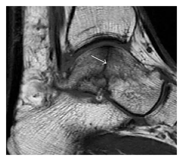
- Cartilage and the talar dome: the cartilage and bone beneath it on top of the talus are checked for an osteochondral lesion, a small patch of damaged cartilage and bone and a classic reason for an ankle that stays sore after a sprain, and for thinning that can progress to osteoarthritis
- Bone and marrow: bruise-like marrow edema, stress reactions and fractures invisible on X-ray, in the talus, heel bone, navicular, tibia and fibula, and less commonly osteonecrosis of the talus
- The joint and the soft tissue around it: fluid, inflamed synovium, bone spurs, loose bodies, the soft-tissue impingement behind front or back ankle pain, the sinus tarsi (a small canal beneath the ankle), and ganglion cysts or other masses, including in the tarsal tunnel
Two honest limits are worth stating. A standard ankle MRI covers the ankle and hindfoot, so a midfoot or forefoot problem, such as a Lisfranc injury or a Morton's neuroma, needs a dedicated foot MRI, and a report on an ankle study cannot speak to it. And the scan is taken lying down, so it shows structure, not how the ankle behaves under load: whether the syndesmosis widens on standing is better judged on weight-bearing X-rays or CT, which also shows fine fracture detail better than MRI. A careful ankle MRI interpretation says so directly rather than implying the scan settled a question it was never built to answer.
How DocOrbit reads your ankle MRI
Upload your scan for a second opinion (the DICOM files from the study, or a copy of the disc you were given) and a board-certified musculoskeletal radiologist carries out a full ankle MRI second read. They go through the same structures one at a time, from the lateral ligaments and peroneal tendons to the talar cartilage and bone marrow, and write a structured report you can hand to your own doctor or surgeon. The signed report comes back in 24-48 hours. It does not say the first read was wrong; it says what a fresh, focused look at your ankle found, in the clinical language your physician already speaks.
Get your ankle MRI re-read by a subspecialist
Ask the radiologist questions afterward
One thing that surprises most patients: the second opinion does not end when the report arrives. You can ask a radiologist online, messaging the musculoskeletal radiologist directly about a phrase in the report, whether a finding really explains your pain, or what to ask your orthopedic surgeon next, and get a written answer back. A second opinion on an ankle MRI is built around that conversation rather than treating the report as the last word.
Or get an answer in minutes with Orbius
If you do not want to wait 24-48 hours for a signed report, DocOrbit also offers Orbius, an AI radiology agent built for exactly this kind of online radiology second opinion. It reads the same ankle MRI and returns a structured report, findings, conclusion and suggested next steps, in minutes, at a lower cost, at any hour. It is an honest trade: speed and price against a human subspecialist's training and signature. The two are separate choices, not two steps of one service: if you want a radiologist afterwards, you can book one in a click with your scans carried over, but that is a separate consultation in which the radiologist reads the scan fresh and writes an independent report rather than reviewing or signing Orbius's.
How do I send my ankle MRI for a second opinion?
Upload the DICOM files from your scan, or the CD you were given at the imaging center, directly through the DocOrbit platform. Nothing needs to be mailed or dropped off anywhere, the images go straight to the radiologist reviewing your case.
Do I need the actual MRI images, or is the written report enough?
You need the images themselves, not just the written report. A genuine second opinion means a radiologist looks at your ankle MRI directly, the way the first one did, rather than reviewing someone else's description.
Will my orthopedic surgeon accept a second opinion report?
Yes. The report follows the same structured format radiologists use everywhere, with findings and an impression your orthopedic surgeon or primary doctor can read and act on like any other radiology report, and you are free to share it directly.
How long does an ankle MRI second opinion take?
A signed report from a board-certified musculoskeletal radiologist typically comes back in 24 to 48 hours. If you want an answer sooner, Orbius, DocOrbit's AI radiology agent, returns a structured report on the same images in minutes.
Is it worth getting a second opinion before ankle surgery, or if my MRI was reported as normal?
Both are common reasons. Before a scheduled operation, a second read confirms the imaging genuinely supports the plan; if the scan came back normal but the ankle still hurts, a fresh, unhurried review sometimes picks up a subtle finding, such as a thin tendon split or a small cartilage defect, that a fast first read was not looking for, or confirms that normal really does mean normal.
Key takeaways
- An ankle MRI runs to well over a hundred images of ligaments, tendons, cartilage and bone, more than one fast read is built to comb
- An MSK radiologist reads joints, tendons and bone daily and is more likely than a generalist to catch a thin peroneal tendon split or a talar cartilage defect
- A proper review walks the anatomy structure by structure and says plainly what an ankle MRI cannot show: the midfoot, the forefoot and behavior under load
- DocOrbit's signed MSK radiologist read takes 24-48 hours; Orbius, the AI option, returns a structured report in minutes
- You can message the radiologist with questions after the report arrives
Can I get a second opinion on my ankle MRI online?
Yes. You upload your images from home, with no CD to post and no second trip, and a subspecialist radiologist re-reads the study and writes a structured report you can hand to your own doctor within 24 to 48 hours. You can message the radiologist with questions about the scan and what the report means for you, and Orbius is there if you would rather have a report in minutes.
This article is for general information only and is not medical advice. Always discuss your imaging results and any next steps with a qualified physician.
