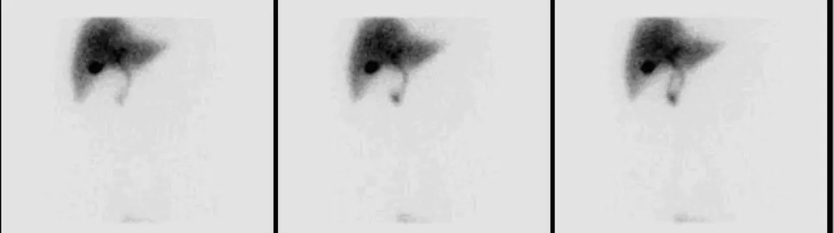
You have had episodes of pain under the right ribs, an ultrasound that came back clean, and now a scan with an unfamiliar name has produced a report full of words like tracer, visualisation, and ejection fraction. Here is the useful framing before anything else: a HIDA scan is not a hunt for a mass. It is a functional test, ordered because your doctor already suspects the gallbladder or the bile ducts and wants to watch them work rather than simply look at them.
What a HIDA scan actually is
HIDA stands for hepatobiliary iminodiacetic acid, the family of tracers used for the test. You will also see it written as hepatobiliary scintigraphy or a cholescintigram, and all three names describe the same study.
A small amount of a radioactive tracer is injected into a vein. Your liver treats it almost exactly like bile, extracting it from the blood and pushing it out through the bile ducts. A gamma camera sitting above your abdomen records where the tracer goes, minute by minute, for about an hour.
That sequence is the whole point. An ultrasound gives one beautiful still picture of anatomy. A HIDA scan gives a film of plumbing: liver, then ducts, then gallbladder, then small intestine. If the tracer stalls somewhere along that path, the images show exactly where.
Why your doctor ordered it
- Typical biliary pain with a normal ultrasound. The single most common reason. Real attacks, no stones on the scan.
- Suspected acute cholecystitis. When the clinical picture suggests an inflamed, blocked gallbladder but the ultrasound is equivocal.
- Suspected biliary dyskinesia. A gallbladder that squeezes poorly even though nothing is obstructing it.
- A suspected bile leak after gallbladder surgery or liver trauma, where tracer appears outside the ducts.
- Unexplained jaundice in a newborn, where the study helps separate biliary atresia from other causes.
If your ultrasound did show stones, you generally do not need this test. Stones plus classic symptoms is usually enough, which is why gallstones found on ultrasound tend to go straight to a surgical conversation instead.
What the appointment feels like
You will be asked to fast for at least four hours, because a gallbladder that has just emptied after a meal cannot fill with tracer and the study becomes uninterpretable. Very long fasting or feeding through a drip causes the opposite problem, a sludge-filled gallbladder that refuses to fill for reasons unrelated to disease, so tell the department if either applies to you.
After the injection you lie still while the camera records. Roughly an hour in, many centres give a second drug called cholecystokinin, or CCK, which is the same hormone your small intestine releases after a fatty meal. It tells the gallbladder to contract.
This is the part patients remember. CCK can produce nausea, cramping, or a wave of the exact pain that brought you to the doctor. Unpleasant as that is, reproducing your symptoms during a measured contraction is genuinely informative, and it is worth telling the technologist what you feel and when.
Reading the report: filling, timing, and the ejection fraction
Three findings carry most of the weight.
Does the gallbladder fill? Normally tracer appears in it within an hour. If the liver and ducts light up but the gallbladder never does, the report will say non-visualisation, and the usual explanation is a blocked cystic duct. Paired with fever and tenderness, that is the classic signature of acute cholecystitis.
Does tracer reach the bowel? Tracer that never arrives in the small intestine suggests obstruction further down the common bile duct.
How well does the gallbladder squeeze? This is the ejection fraction, the percentage of tracer expelled after CCK. Most centres use a threshold somewhere between 35 and 40 percent. Below that, the report may raise biliary dyskinesia, sometimes described as a functional gallbladder disorder.
One caveat is worth knowing, because it changes how much weight the number deserves. The result depends heavily on how the CCK was administered. A slow infusion over 30 to 60 minutes is far more reproducible than a fast injection, which can force an artificial contraction and drive the number down. A borderline value from a rapid protocol is a much softer finding than the same number from a slow one.
What happens next
For non-visualisation with acute symptoms, the pathway is usually antibiotics and surgery, sometimes urgently. For a low ejection fraction without infection, the conversation is more nuanced.
Gallbladder removal helps a substantial share of people with a low ejection fraction and textbook biliary attacks, meaning severe pain in the right upper abdomen or the pit of the stomach, often radiating to the right shoulder blade, lasting at least 30 minutes and settling in between. The picture is far less predictable when the dominant complaints are bloating, heartburn, or grumbling daily discomfort, because those overlap heavily with reflux, gastritis, and irritable bowel. Surgery in that group leads to a good number of people who feel no better afterwards.
A normal study is meaningful too. It pushes the search towards the stomach, the pancreas, or a functional gut disorder, and that redirection can save months of chasing the wrong organ.
Things that genuinely help while you wait
- Smaller, lower-fat meals. Large fatty meals are the classic trigger for an attack.
- Steady, gradual weight loss if you are carrying extra weight. Crash dieting raises the risk of stone formation.
- Keeping a short pain diary with what you ate, when the pain started, how long it lasted, and where it went. Duration and pattern are what a surgeon weighs most heavily.
- Noting fever, persistent vomiting, or yellowing of the eyes. Those move the situation from an outpatient question to a same-day one.
Why a second read can help
Functional nuclear medicine studies leave more room for interpretation than most patients expect. The ejection fraction depends on protocol, the software's region of interest, and how the images are drawn, and non-visualisation on delayed pictures can be read differently by different eyes. If your report is borderline, or if a low number is about to become the reason for surgery, an independent radiology read is a reasonable step. DocOrbit lets you upload the study and receive an expert second opinion you can bring back to your own doctor, in the same spirit as knowing when to ask for a second radiological opinion for any imaging result.
What is a normal gallbladder ejection fraction?
Most centres treat an ejection fraction above 35 to 40 percent as normal, though the exact cut-off depends on the protocol and how the CCK was given. A slow infusion over 30 to 60 minutes gives a far more reliable number than a rapid injection, which can squeeze the gallbladder artificially and produce a falsely low result. This is one reason two labs can report different numbers on the same person.
Does a HIDA scan hurt?
The scan itself is painless. You lie still on a table with a camera above your abdomen while a small dose of radioactive tracer travels from your bloodstream into your liver and bile. The one part people do notice is the CCK phase, which can bring on nausea, cramping, or the same right-sided pain that brought you in. That reaction is uncomfortable but it is also useful information for your doctor.
How long does a HIDA scan take?
Plan for one to two hours. Imaging usually runs for about an hour after the tracer is injected, and if CCK is given to measure the ejection fraction, that adds another 30 to 60 minutes. If the gallbladder has not appeared by the end of the standard imaging window, delayed pictures may be taken up to about four hours, which makes the appointment longer.
What does it mean if my gallbladder did not show up on the HIDA scan?
Non-visualisation means tracer reached the liver and the bile ducts but never entered the gallbladder, which usually points to a blocked cystic duct. In someone with fever and severe right upper abdominal pain, that is the classic picture of acute cholecystitis. In other settings it can reflect chronic scarring, recent fasting or prolonged intravenous feeding, so it is always read alongside your symptoms and blood tests.
Is the radiation from a HIDA scan dangerous?
The dose is modest, broadly comparable to a CT scan of the abdomen and delivered by a tracer that clears from the body within about a day. For an adult being investigated for repeated biliary pain, the information gained generally outweighs that exposure. Tell the team if you are pregnant or breastfeeding, since the timing and the protocol may be adjusted.
Will surgery fix my pain if the ejection fraction is low?
Many people with a low ejection fraction and typical biliary pain do improve after gallbladder removal, but the evidence is less clear-cut than it is for gallstones. Results are best when the pain pattern is classic, meaning severe episodes in the right upper abdomen lasting at least half an hour, and less predictable when the main complaints are bloating, heartburn, or daily discomfort. A good surgeon will talk through that difference before booking anything.
Key takeaways
- A HIDA scan watches the gallbladder and bile ducts work, which is a different question from what an ultrasound answers.
- Non-visualisation of the gallbladder in someone acutely unwell usually means a blocked cystic duct and acute cholecystitis.
- The ejection fraction is protocol-dependent; a slow CCK infusion gives a far more trustworthy number than a rapid injection.
- A low number matters most when the pain pattern is classic biliary colic rather than bloating or daily discomfort.
- A normal study is useful information, redirecting the search towards the stomach, pancreas, or gut.
If the rest of your report reads like another language, our free radiology report explainer rewrites it in everyday words, defines the terms it uses and suggests what to ask at your next appointment.
Can I get a second opinion on my nuclear medicine scan online?
Yes. Online radiology second opinions run entirely from home: you upload your images, with no CD to post and no second trip, and a subspecialist radiologist re-reads the study and writes a structured report you can give to your own doctor. DocOrbit returns that signed second read in 24 to 48 hours. You can then message the radiologist with questions about the nuclear medicine scan and about what the report actually means for you.
This article is for general information only and is not medical advice. Always discuss your imaging results and any next steps with a qualified physician.