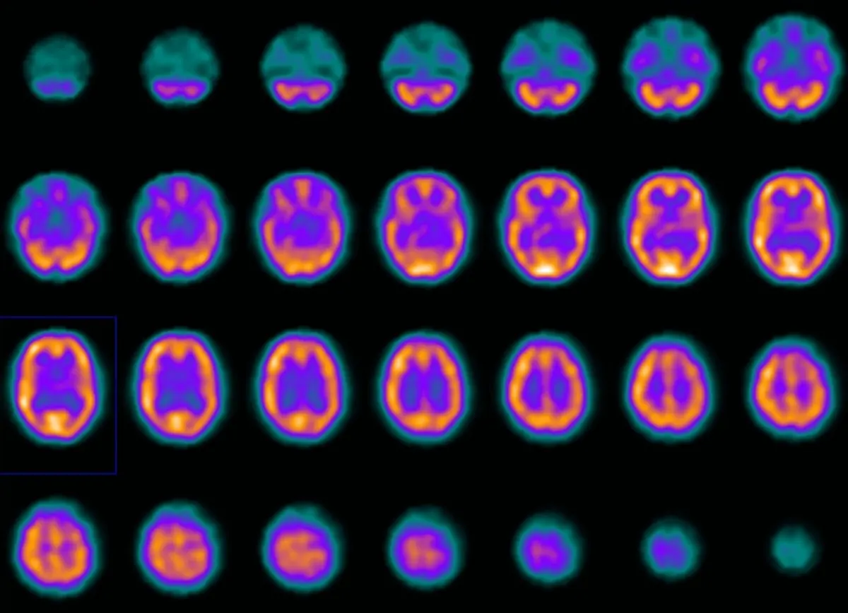
You have had a tremor, or slowness, or a change in your handwriting, and after months of not-quite-answers somebody finally ordered a DaTscan. Then came the iodine drops, the injection, the long wait, the camera, and now a report that talks about reduced uptake in the putamen or symmetric striatal binding. Here is the honest framing before anything else: this scan was ordered because your picture was not clear-cut, and its job is narrow. It answers one question well, and deliberately leaves several others to your neurologist.
What a DaTscan actually measures
Deep in the brain sits a small structure called the striatum, made up of the caudate and the putamen. Nerve endings arriving there from the substantia nigra carry dopamine, the chemical that lets movement start smoothly and stop cleanly. On the surface of those nerve endings are dopamine transporters, tiny recycling pumps.
A DaTscan uses a radioactive tracer, ioflupane, that sticks to those transporters. A gamma camera then maps where the tracer collected. More tracer means more surviving dopamine nerve endings; less tracer means fewer.
So the scan is not looking at Parkinson's disease directly, and it is not looking at the brain's structure at all. It is counting a specific population of nerve endings. Everything a DaTscan can and cannot tell you follows from that one fact.
Why your neurologist ordered it
Parkinson's disease is diagnosed clinically, by examination and history, and an experienced neurologist is right most of the time without any scan. A DaTscan gets added when the picture is genuinely ambiguous, most often in these situations:
- Tremor that could be essential tremor. Essential tremor is common, often runs in families, and can look convincingly like early Parkinson's, but the dopamine system is intact in it.
- Symptoms that started after a new medication. Some anti-nausea and antipsychotic drugs block dopamine receptors and produce a very good imitation of Parkinson's that reverses when the drug is stopped.
- An unclear response to levodopa. When a trial of treatment helps less than expected, the question becomes whether the dopamine system was ever the problem.
- Dementia with an uncertain cause. The scan is also used to help separate dementia with Lewy bodies from Alzheimer's disease, because the first affects the dopamine system and the second does not.
- Vascular or functional parkinsonism. Slowness can also come from small vessel disease in the brain, the kind described in our article on chronic ischemic changes on MRI, or from a functional movement disorder.
In other words, the scan is usually ordered to rule something out rather than to confirm a diagnosis your team has already made.
What a normal scan looks like
On a normal study, the tracer collects densely and symmetrically in the striatum on both sides, producing two crescent shapes that radiologists often describe as comma-shaped. The signal fills the head of the caudate at the front and runs back through the putamen.
A normal result means the dopamine nerve endings are intact. That makes Parkinson's disease, multiple system atrophy, progressive supranuclear palsy, and dementia with Lewy bodies unlikely, and it redirects attention to the conditions that do not destroy those cells: essential tremor, drug-induced parkinsonism, vascular parkinsonism, and functional movement disorders. For many people this is the moment a long diagnostic detour finally ends.
What an abnormal scan looks like
When dopamine nerve endings have been lost, the tracer signal shrinks from the back forward. The putamen fades first, so the comma shortens into something closer to a full stop or a small oval, and the loss is often clearly worse on one side, matching whichever side of the body is more affected.
An abnormal scan tells your neurologist that this is a degenerative dopaminergic condition rather than a mimic. It does not tell them which one. And this is where expectations and reality most often part company.
What a DaTscan cannot tell you
- Which parkinsonian condition you have. Parkinson's disease and the atypical syndromes reduce the same signal. Telling them apart is a clinical job, sometimes helped by other imaging.
- How severe your disease is. The degree of reduction correlates only loosely with symptoms. People with similar-looking scans can be at quite different stages.
- How fast things will progress. The scan is a snapshot of the dopamine system now, not a forecast.
- Whether treatment is working. Symptoms and function answer that question, not repeat imaging. DaTscans are rarely repeated for that reason.
Reports are written by eye and often supported by semi-quantitative numbers comparing striatal uptake with a background region. Those numbers help, but they are influenced by the camera, the software, and the reference values used, so they are guides rather than verdicts.
What the day itself involves
You will be asked to take iodine drops or tablets an hour or so before the injection, which protects the thyroid gland from the small amount of free iodine that the tracer releases. The injection itself goes into a vein in the arm and takes seconds. Then there is a wait of roughly three to six hours while the tracer distributes and clears from tissue where it is not wanted. Bring something to read.
The imaging takes about thirty to forty-five minutes, lying on your back with your head gently supported while the camera rotates slowly around it. There is no tunnel and no loud noise, so it is generally easier than an MRI for people who find enclosed spaces difficult. Tremor during the scan can blur the images, so tell the technologist if staying still is hard for you; departments have ways of helping.
A few medicines can interfere with tracer binding, including some antidepressants, stimulants prescribed for attention disorders, certain decongestants, and some anaesthetic agents. Your department sends a list. Standard Parkinson's medicines such as levodopa are usually continued, and no one should stop a prescribed drug without confirming it first.
Why a second read can help
DaTscan reporting rests on pattern recognition, and the difficult cases, mild asymmetries, borderline putaminal loss, or images degraded by movement, are exactly the ones that change what happens next in a person's life. A diagnosis of Parkinson's disease reshapes work, driving, and treatment plans, so it is reasonable to want the images looked at twice. An independent second read through DocOrbit gives you a nuclear medicine opinion in plain language that you can take back to your own neurologist, which is worth considering whenever a scan result and a clinical picture do not sit comfortably together. Our piece on second opinions in neurology covers when that is most useful.
Does a normal DaTscan rule out Parkinson's disease?
A normal scan makes Parkinson's disease and the related degenerative conditions very unlikely, which is why the test is so useful when the diagnosis is uncertain. It points instead toward causes that do not damage dopamine-producing cells, such as essential tremor, a medication side effect, or a functional movement disorder. It is not a certificate that nothing will ever change, so if your symptoms progress in an unexpected way your neurologist may reassess.
Can a DaTscan tell the difference between Parkinson's and other parkinsonian conditions?
No, and this is the most common misunderstanding about the test. Parkinson's disease, multiple system atrophy, progressive supranuclear palsy, and dementia with Lewy bodies all reduce the same dopamine transporter signal, so they can look alike on the scan. The DaTscan tells your neurologist whether the dopamine system is damaged at all; deciding which condition it is remains a clinical judgement based on your examination, your symptoms, and how things evolve.
Do I need to stop my Parkinson's medication before a DaTscan?
Usually not. Levodopa and most standard Parkinson's medicines do not meaningfully change the result, so they are generally continued. What can interfere are certain other drugs, including some antidepressants, stimulants used for attention disorders, some decongestants, and a few anaesthetic agents. The nuclear medicine department will send you a specific list, and you should never stop a prescribed medicine on your own initiative before checking with them.
How much radiation is in a DaTscan?
The dose is in the same broad range as a CT scan of the abdomen, or roughly a couple of years of the natural background radiation everyone receives. You are also given iodine drops or tablets beforehand to protect the thyroid from the small amount of free iodine in the tracer. For an adult being investigated for a movement disorder, that exposure is generally considered a reasonable trade for a clearer diagnosis.
How long does a DaTscan take and when do results come back?
Expect to be at the hospital for most of a morning or afternoon. You take the thyroid-blocking iodine first, receive the tracer injection, then wait around three to six hours before lying still under the camera for roughly thirty to forty-five minutes. The images are usually reported within a few days, and your neurologist will normally discuss them with you at a follow-up appointment rather than by phone.
Key takeaways
- A DaTscan maps surviving dopamine nerve endings in the striatum; it does not image brain structure and does not diagnose a specific disease.
- A normal scan strongly points away from Parkinson's and toward mimics such as essential tremor or drug-induced parkinsonism.
- An abnormal scan confirms dopamine loss but cannot separate Parkinson's disease from the atypical parkinsonian syndromes.
- Severity and prognosis are judged clinically, which is why the scan is rarely repeated.
- Most standard Parkinson's medicines are continued, but some other drugs interfere, so follow the department's list exactly.
Still unsure what the rest of your report says? Paste it into our free radiology report explainer and read it back in plain English, with each medical term defined and a short list of questions worth asking your doctor.
How do I have my nuclear medicine scan reviewed by another radiologist online?
Yes. Online radiology second opinions run entirely from home: you upload your images, with no CD to post and no second trip, and a subspecialist radiologist re-reads the study and writes a structured report you can give to your own doctor. DocOrbit returns that signed second read in 24 to 48 hours. You can then message the radiologist with questions about the nuclear medicine scan and about what the report actually means for you.
This article is for general information only and is not medical advice. Always discuss your imaging results and any next steps with a qualified physician.