If your report mentions "fatty liver," "hepatic steatosis," or "increased liver echogenicity," it is one of the most common findings on abdominal imaging, and in the large majority of patients it is reversible. This guide explains what fatty liver actually is, why MRI gives the clearest picture, what mild, moderate, and severe mean in practice, and what really changes the trajectory.
What fatty liver actually is
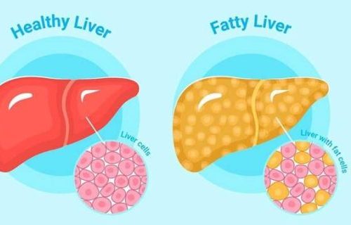
A healthy liver contains a small amount of fat, under about 5% of its weight. When that percentage rises, the result is hepatic steatosis, or fatty liver. Most cases fall under what doctors now call metabolic dysfunction-associated steatotic liver disease (MASLD), which is closely linked to weight, insulin resistance, type 2 diabetes, and high triglycerides. Alcohol is a separate driver of fatty change and is sometimes layered on top.
The condition is unusually common. It is the most frequent chronic liver condition worldwide, and patients are often surprised to see it on a report after an ultrasound done for an unrelated reason. The presence of fat alone is rarely an emergency. What matters is the trajectory and whether inflammation and scarring are joining the picture.
What MRI shows that other tests miss
Three imaging tests are commonly used for fatty liver, and they are not equivalent.
- Ultrasound: fast, widely available, and the usual first test. It describes the liver as mildly, moderately, or severely "bright" (echogenic). It is excellent for screening but cannot give a number, and its accuracy drops in patients with obesity, where image quality is harder.
- CT: it can detect steatosis, but it exposes the patient to radiation dose, so it is not the test of choice when MRI is available.
- MRI: the most accurate non-invasive method. A specific technique called MRI proton density fat fraction (MRI-PDFF) measures the percentage of fat in the liver directly. A result of "11.3%" is much more actionable than "moderate steatosis."
MRI also has the advantage of being repeatable: a second MRI six or twelve months later can show whether your liver fat is dropping, holding steady, or rising. That picture stays objective, regardless of how you feel or what your weight is doing.
Mild, moderate, and severe: what the numbers mean
There is no single universally agreed boundary, but the practical ranges used in MRI-PDFF research are roughly:
- Normal: less than about 5% liver fat.
- Mild steatosis: roughly 5% to 15%.
- Moderate steatosis: roughly 15% to 25%.
- Severe steatosis: above about 25%, sometimes much higher.
The category itself is less important than the direction of change. A patient who drops from 18% to 9% over a year is doing well, even if the radiologist still uses the word "steatosis." A patient whose fat fraction rises from 8% to 17% needs a closer look at lifestyle and metabolic factors, even if the absolute number is not alarming.
When fatty liver matters and when it is incidental
Most fatty liver does not cause symptoms. Patients usually feel completely well. The clinical concern is not the fat itself but its potential to evolve:
- Steatosis alone: fat in the liver without significant inflammation. Usually reversible with lifestyle change.
- MASH (formerly NASH): fat plus inflammation and liver-cell injury. Over years, this can lead to fibrosis (scarring).
- Fibrosis: accumulating scar tissue. Mild fibrosis is reversible; advanced fibrosis is harder to reverse.
- Cirrhosis: advanced scarring with structural distortion. A long-term endpoint of progressive disease, not a near-term consequence of mild fatty liver.
MRI elastography or transient elastography (FibroScan) is used to estimate fibrosis non-invasively, and blood-based scores (FIB-4, APRI) add information. The MRI fat fraction does not tell you about scarring on its own. These are different questions, answered by different tests.
What actually moves the needle
Almost everything that helps fatty liver is a lifestyle intervention. The evidence here is unusually strong:
- Weight loss of about 5–10% of body weight: the single most reliable intervention. Liver fat often drops sharply at this threshold.
- Reduce ultra-processed food, refined sugars, and sugary drinks: fructose in particular drives liver fat synthesis.
- Increase physical activity: both aerobic exercise and resistance training reduce liver fat independent of weight loss.
- Limit or stop alcohol: even moderate alcohol intake can add to existing steatosis.
- Treat diabetes, prediabetes, and high triglycerides: these conditions share the same metabolic root as fatty liver and respond to similar levers.
- Coffee, in normal amounts: observational data consistently associates regular coffee intake with lower fibrosis risk.
Medications specifically for MASH (resmetirom, GLP-1 receptor agonists in selected patients) are an emerging area, but lifestyle remains the foundation of treatment for almost everyone.
Why a second read can help
Liver fat quantification is one of the most reader-dependent steps in abdominal imaging. Whether the right MRI sequence was used, whether the measurement was placed on representative liver tissue, and whether other findings (focal fat, focal sparing, masses) were correctly characterized all matter. DocOrbit can provide an expert second-opinion radiology report that you can share with your treating physician. It includes a careful look at the fat fraction and any associated findings. For broader context on when a second imaging read is worth the effort, see the essential role of second opinions in radiology.
Common pitfalls in liver MRI interpretation
A few specific situations are worth knowing about, because they tend to trip up generalist reads of an abdominal MRI:
- Focal fat and focal sparing: fat does not always distribute evenly. Patches of higher or lower fat content next to gallbladder fossa or portal vein branches can mimic a mass on ultrasound or CT, and MRI is usually the test that resolves the question.
- Iron overload: co-existing iron deposition (hemochromatosis, transfusion-related) changes the MRI signal and, if not accounted for, can artificially raise or lower the measured fat fraction. A proper protocol corrects for this.
- Mass lesions in a fatty liver: a fatty background makes some lesions easier to see and others harder. A second read can confirm that no nodule or hemangioma was overlooked.
- Wrong-sequence reporting: a fat fraction taken from a standard chemical-shift sequence is not the same as a true MRI-PDFF. The report should specify which technique was used.
None of these are reasons to distrust a single read. They are reasons the same MRI, looked at by a second reader with the right protocol in mind, sometimes yields a more accurate number or a better-characterized incidental finding.
What to expect at follow-up
If your first MRI shows fatty liver, what happens next depends on the fat fraction, your other risk factors, and whether there is any sign of fibrosis. A typical pattern looks like this:
- Confirmation that other liver diseases are excluded: viral hepatitis, autoimmune liver disease, and iron overload have to be ruled out with blood tests.
- Fibrosis assessment: usually MRI elastography, transient elastography (FibroScan), or a blood-based score. This determines whether the focus is lifestyle alone or whether you need closer follow-up.
- Repeat imaging in 6–12 months: for patients with significant steatosis or risk factors, a follow-up MRI shows whether the fat fraction is moving in the right direction.
- Long-term metabolic care: regular blood work for liver function, lipids, and glucose, coordinated with your primary care doctor or hepatologist.
What does fatty liver on MRI actually mean?
It means the liver contains more fat than normal. Specifically, more than about 5% of liver weight is fat. On MRI, this can be measured directly as a percentage using a technique called MRI-PDFF (proton density fat fraction). Unlike ultrasound, which describes the liver in broad categories, MRI-PDFF gives a specific number that can be tracked over time as you make lifestyle changes.
Is fatty liver dangerous?
Most fatty liver is mild and reversible, especially when caught early and addressed with diet, exercise, and weight management. The concern is when fat is combined with inflammation and scarring. This more advanced form is called MASH (formerly NASH). That can progress to fibrosis and, over many years, cirrhosis. The MRI fat percentage alone does not tell you which group you are in; that needs additional testing and clinical correlation.
Why is MRI better than ultrasound for fatty liver?
Ultrasound is a useful screening tool, but it only sorts fatty liver into broad grades (mild, moderate, severe) based on how bright the liver looks. MRI-PDFF reports an actual percentage, for example 12.4% liver fat, and is sensitive to small changes over time. This makes MRI especially useful for tracking whether your liver is improving with diet, exercise, or treatment.
Can fatty liver be reversed?
Yes, in most cases. Sustained weight loss of around 5–10% of body weight has been shown to reduce liver fat substantially. Cutting back on alcohol, processed sugars, and ultra-processed foods, increasing physical activity, and treating diabetes and high cholesterol all help. Improvement is often visible on a follow-up MRI 6–12 months later.
Key takeaways
- Fatty liver is the most common chronic liver condition worldwide and, in early stages, is usually reversible.
- MRI-PDFF gives a specific liver fat percentage and is the most accurate non-invasive way to track change over time.
- The MRI tells you about fat; a separate test (elastography or blood-based scores) is needed to assess scarring.
- Sustained 5–10% weight loss is the single strongest intervention; cutting refined sugars, alcohol, and ultra-processed food adds to it.
- A second imaging read can confirm the fat fraction was measured correctly and that no other findings were missed.
This article is for general information only and is not medical advice. Always discuss your imaging results and any next steps with a qualified physician.