Seeing the words "pleural effusion" on a chest X-ray or CT report can be unsettling, especially if you went in for something unrelated. Here is the reassuring starting point: a pleural effusion simply means a little extra fluid has collected around the lung, and in many cases it is a temporary, treatable response to something else going on in the body. It is a finding, not a diagnosis on its own. This article explains what the term means, what commonly causes it, when it matters, and how doctors usually handle it.
What does "pleural effusion" mean?
Your lungs are wrapped in two thin layers of tissue called the pleura. Between them is a tiny amount of lubricating fluid that lets the lung glide smoothly as you breathe. Normally it amounts to just a few teaspoons. A pleural effusion is when more fluid than normal builds up in that space.
On imaging, radiologists describe it in a few ways:
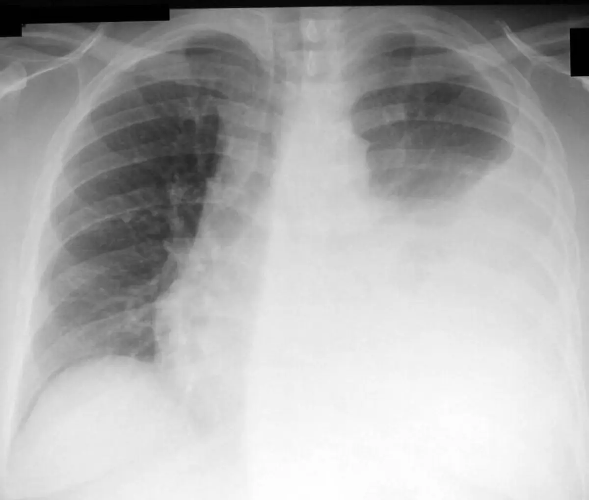
- On a chest X-ray, fluid often shows up as a white area at the bottom of one lung, sometimes blunting the sharp angle where the lung meets the diaphragm
- On a CT scan, the fluid is seen more precisely, and its density can hint at whether it is simple watery fluid, blood, or something thicker
- Reports may call it "small", "moderate", or "large", and note whether it is on one side (unilateral) or both (bilateral)
Whether the effusion is on one side or both is an important clue. Fluid on both sides more often reflects a body-wide issue like heart failure, while fluid on just one side raises a wider list of local possibilities.
Common causes
Pleural effusions are grouped by the type of fluid, which points toward the cause. "Transudates" are thin, watery fluid usually from pressure or fluid-balance problems, while "exudates" are protein-rich fluid usually from inflammation, infection, or irritation of the pleura.
- Heart failure: the most common cause overall, often producing fluid on both sides
- Pneumonia or chest infection: inflammation can pull fluid into the pleural space
- Recent surgery or procedures: fluid is especially common after abdominal or heart surgery
- Liver or kidney disease: these can lead the body to retain fluid
- Pulmonary embolism: a blood clot in the lung
- Cancer: a less common but important cause, usually investigated when the fluid is unexplained
If your report also mentions other chest findings, it can help to read about how individual abnormalities are interpreted. For example, our explainer on a lung nodule found on a CT scan walks through how a single finding is put into context rather than read in isolation.
Is it serious?
The honest answer is that a pleural effusion can range from trivial to important, and the amount of fluid is only part of the story. A small effusion after surgery or with a passing infection is often a minor, self-limited thing. A large or steadily growing effusion, or one with no obvious explanation, is taken more seriously and worked up further.
Doctors pay particular attention when an effusion comes with fever, weight loss, coughing up blood, or significant breathlessness. These symptoms point toward a cause that needs prompt evaluation. As with most imaging findings, interpretation depends on your symptoms, exam, blood tests, and history, not the scan alone.
Symptoms
Many small effusions cause no symptoms and are picked up incidentally. When symptoms do appear, they usually come from the fluid pressing on the lung:
- Shortness of breath, especially when lying flat or with exertion
- A feeling of heaviness or pressure in the chest
- A dry cough
- Chest discomfort that may sharpen with a deep breath
How strong the symptoms are depends largely on how much fluid is present and how quickly it accumulated. A small amount that built up slowly may go unnoticed, while a rapid buildup can feel much more pronounced.
How is it diagnosed and followed up?
An effusion is often first seen on a chest X-ray, then better characterized with ultrasound or CT. When the cause is not obvious, the key step is usually a procedure called thoracentesis, in which a doctor samples the fluid with a thin needle so it can be analyzed in the lab. That single test answers many questions at once: it shows whether the fluid is transudate or exudate, looks for infection, and checks for abnormal cells.
Follow-up cadence varies by patient. A small effusion with a clear, treatable cause may simply be rechecked once the underlying problem is treated, while an unexplained one usually prompts more focused testing.
Treatment options
Treatment almost always starts with the underlying cause rather than the fluid itself:
- Heart failure: diuretics and heart-failure management often reduce the fluid
- Infection: antibiotics, sometimes with drainage if the fluid is infected
- Large, symptomatic effusions: these are drained with thoracentesis to relieve breathlessness and obtain a sample
- Recurrent effusions: these may need a longer-term drain or a procedure to prevent the fluid from coming back
Many effusions need no aggressive treatment at all once the root cause is addressed. The goal is to relieve symptoms and resolve the condition driving the fluid.
Why a second read can help
Because a pleural effusion is a clue rather than an answer, the value of imaging lies in how carefully it is interpreted alongside everything else about you. A clear, expert read can help distinguish a routine effusion from one that deserves a closer look, and it gives you something concrete to discuss with your own doctor. DocOrbit offers an expert second read of your scan that you can share directly with your care team. It is a way to feel confident the finding has been understood in full context. If you are weighing whether to seek one, our piece on when to get a second radiological opinion may help you decide, and you can read more broadly about the role of second opinions in radiology.
Is a pleural effusion serious?
It depends entirely on what is causing it. A small effusion from a chest infection or recent surgery is often temporary and resolves with treatment of the underlying problem. A larger or recurring effusion can point to a condition that needs closer attention, such as heart failure or, less commonly, cancer. The fluid itself is a clue, not a diagnosis, so your doctor will look at your symptoms, exam, and other tests to judge how concerned to be.
What does a pleural effusion feel like?
Small effusions often cause no symptoms at all and are found by chance on a scan. Larger ones can cause shortness of breath, a feeling of chest heaviness or pressure, and sometimes a dry cough or pain that worsens with a deep breath. Symptoms usually relate to how much fluid is present and how quickly it built up.
How is fluid around the lung treated?
Treatment targets the cause first. An effusion from heart failure may shrink with diuretics, and one from infection improves with antibiotics. If a large effusion is making it hard to breathe, a doctor can drain it with a needle in a quick procedure called thoracentesis, which also lets the fluid be tested in the lab. Recurring effusions sometimes need a longer-term drain or other procedures.
Does a pleural effusion always mean cancer?
No. Most pleural effusions are caused by non-cancerous conditions such as heart failure, pneumonia, or fluid shifts after surgery. Cancer is one possible cause, but it is far from the most common one. The fluid is usually sampled and analyzed when the cause is unclear, which helps separate benign causes from ones that need more workup.
Can a pleural effusion go away on its own?
Yes, many do, especially small effusions tied to a temporary problem like a viral infection or recent surgery. As the underlying issue settles, the body reabsorbs the fluid. Larger effusions or those from a chronic condition are less likely to clear without treating the root cause.
Key takeaways
- A pleural effusion means extra fluid has collected around the lung. It is a finding, not a diagnosis on its own
- The most common cause is heart failure, followed by infection and post-surgical fluid shifts; cancer is a less common cause
- Small effusions often cause no symptoms; larger ones can cause breathlessness and chest pressure
- Treatment focuses on the underlying cause, with drainage available when the fluid causes symptoms or needs testing
- Interpretation always depends on your symptoms, exam, and history rather than the scan alone
This article is for general information only and is not medical advice. Always discuss your imaging results and any next steps with a qualified physician.