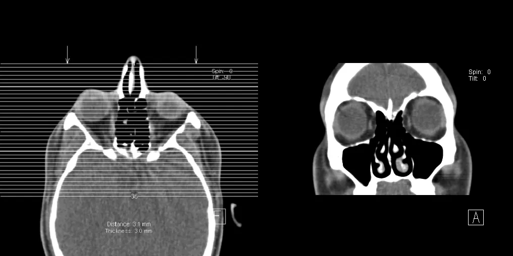
If a CT or MRI report mentions "mucosal thickening" of the paranasal sinuses, it is one of the most common phrases head imaging ever produces. On its own, it is rarely something to worry about. It usually means the soft tissue lining your sinuses looks a little swollen, often from something as ordinary as a recent cold or seasonal allergies. This guide explains what the phrase means, what causes it, and when it actually matters.
What "mucosal thickening" of the paranasal sinuses means
The paranasal sinuses are air-filled spaces in the bones around your nose and eyes. There are four pairs: the maxillary sinuses (in the cheeks), the ethmoid sinuses (between the eyes), the frontal sinuses (in the forehead), and the sphenoid sinuses (deeper, behind the nose). Each one is lined by a thin, moist layer of tissue called mucosa.
In a healthy sinus, that lining is so thin it is barely visible, and the sinus reads as mostly black because it is filled with air. When the mucosa becomes irritated or inflamed, it swells and holds more fluid, and that thicker, brighter rim is what the radiologist describes as mucosal thickening.
Reports phrase it in several ways:
- Mild mucosal thickening: a thin rim of swollen lining, usually a few millimeters.
- Mucosal thickening of the maxillary/ethmoid sinus: naming which sinus is involved.
- Mucoperiosteal thickening: the same finding, using a more technical term.
- Near-complete or complete opacification: the sinus looks filled rather than air-filled, a more significant degree.
The degree, which sinuses are involved, and whether one or both sides are affected are the details that carry the most weight.
Common causes
Mucosal thickening is a nonspecific finding: many different, mostly benign processes produce the same appearance. The most frequent include:
- A recent or current viral infection: a common cold can keep the sinus lining swollen for days or weeks after you feel better.
- Allergies: allergic rhinitis (hay fever, dust, pet dander) is a leading cause of persistent thickening.
- Acute or chronic sinusitis: inflammation or infection of the sinuses, sometimes bacterial.
- Environmental irritants: cigarette smoke, air pollution, and very dry indoor air.
- Anatomical factors: a deviated nasal septum or nasal polyps that keep sinuses from draining well.
- Dental causes: an infected or recently treated upper back tooth sits close enough to irritate the maxillary sinus floor.
- Recent nasal or sinus surgery: expected post-operative change.
It often turns up in people with no sinus complaints at all. Large imaging studies find it in a substantial share of scans done for unrelated reasons, such as a brain MRI ordered for headache or to check for chronic ischemic changes.
Is it serious?
For the large majority of people, mild mucosal thickening is not serious. It is closer to a normal variant than a disease, in the same family as a small lung nodule on a chest CT: noticed, noted, and usually left alone.
It deserves a closer look when:
- It comes with symptoms: facial pain or pressure, nasal blockage, thick discharge, or a reduced sense of smell, especially when they last more than 12 weeks (a pattern called chronic rhinosinusitis).
- An air-fluid level is described: a flat line of fluid inside the sinus, which can point to acute sinusitis.
- One sinus is completely opacified: particularly on one side only, which sometimes prompts evaluation for a polyp, a fungal cause, or, rarely, a growth.
- The report mentions bone changes: erosion or thinning of the sinus walls is uncommon and warrants specialist review.
One-sided findings are generally taken more seriously than symmetric, both-sided thickening, which is far more often simple inflammation.
Symptoms
Many people with mucosal thickening have no symptoms at all. That is exactly why it so often turns up as an incidental finding. When symptoms are present, they usually reflect sinus inflammation rather than the thickening itself:
- Nasal congestion or a blocked-nose feeling.
- Facial pressure or pain over the cheeks, forehead, or between the eyes.
- Postnasal drip or thick nasal discharge.
- A reduced or altered sense of smell.
- Headache, or a dull ache that worsens when bending forward.
How it is followed up
There is no routine imaging follow-up for mild, incidental mucosal thickening. When there are no sinus symptoms, it is typically just documented.
When symptoms are present, the next steps are clinical rather than radiological:
- A primary care doctor or an ear, nose, and throat (ENT) specialist correlates the scan with your history.
- Acute sinusitis is diagnosed mainly from symptoms; imaging is not required to confirm an uncomplicated case.
- Chronic or recurrent symptoms may lead to a nasal endoscopy (a thin camera passed into the nose) and, in some cases, a dedicated sinus CT for detail or surgical planning.
The imaging describes the lining; your doctor decides whether it explains how you feel.
Treatment and lifestyle
The mucosal thickening itself is not treated. What gets treated is the underlying irritation, and when treatment is needed it is usually straightforward:
- Saline nasal rinses or sprays: simple, safe, and helpful for both allergies and infection.
- Intranasal corticosteroid sprays: a mainstay for allergic and chronic inflammation.
- Allergy management: antihistamines and avoiding known triggers.
- Short-term decongestants: for temporary relief, not long-term use.
- Antibiotics: only when a bacterial sinus infection is genuinely suspected, not for every thickened sinus.
- Treating a dental cause: when maxillary thickening traces back to an upper tooth.
For chronic rhinosinusitis that does not settle with these measures, an ENT specialist may consider functional endoscopic sinus surgery in selected cases. Day to day, not smoking, keeping indoor air from getting too dry, and managing allergies reduce how often the lining flares up.
Why a second read can help
Whether mild mucosal thickening matters depends heavily on how it is read against your symptoms, and that judgment can vary between radiologists. A second read can confirm the degree is genuinely mild and flag whether a one-sided or fully opacified sinus deserves more attention. DocOrbit provides an expert second-opinion radiology report you can share with your own physician. That can be useful when a report's wording leaves you unsure whether a sinus finding is routine or worth a specialist visit.
Is paranasal sinus mucosal thickening serious?
In most cases, no. Mild mucosal thickening is one of the most common incidental findings on head CT and MRI, and on its own it is closer to a normal variant than a disease. It becomes more meaningful when it is paired with symptoms such as facial pain, congestion, or reduced smell, when a sinus is completely opacified, or when only one side is affected. Your doctor weighs the scan against how you actually feel.
Does mucosal thickening always mean I have a sinus infection?
No. Mucosal thickening simply means the sinus lining looks swollen, and many things other than infection cause it. Allergies, a recent cold, smoke or dry air, and a deviated septum are all common culprits. An active bacterial infection is usually suspected from symptoms and findings like an air-fluid level, not from thickening alone. Many people with thickening on a scan have no infection at all.
What does mild mucosal thickening mean?
Mild mucosal thickening describes a thin rim of swollen lining, often just a few millimeters, with the sinus still mostly air-filled. There is no single universal cutoff, and radiologists use the term in the context of the whole scan. Mild, symmetric thickening in someone without sinus symptoms is typically considered nonspecific and not clinically important.
Can paranasal sinus mucosal thickening go away on its own?
Often, yes. When the cause is a passing viral infection or a seasonal allergy flare, the lining frequently returns to normal once the trigger settles. Thickening that comes from ongoing allergies, chronic inflammation, or an anatomical problem tends to persist until that underlying cause is addressed.
Key takeaways
- Mucosal thickening means the lining of one or more paranasal sinuses looks swollen on a CT or MRI.
- It is a very common and usually incidental finding. A recent cold, allergies, or irritants are the usual reasons.
- Mild, symmetric thickening without symptoms generally needs no treatment or follow-up.
- It matters more when symptoms last beyond 12 weeks, when a sinus is fully opacified, or when only one side is involved.
Still unsure what the rest of your report says? Paste it into our free radiology report explainer and read it back in plain English, with each medical term defined and a short list of questions worth asking your doctor.
Can I ask a radiologist about my scan report online?
Yes. Online radiology second opinions run entirely from home: you upload your images, with no CD to post and no second trip, and a subspecialist radiologist re-reads the study and writes a structured report you can give to your own doctor. DocOrbit returns that signed second read in 24 to 48 hours. You can then message the radiologist with questions about the scan and about what the report actually means for you.
This article is for general information only and is not medical advice. Always discuss your imaging results and any next steps with a qualified physician.
