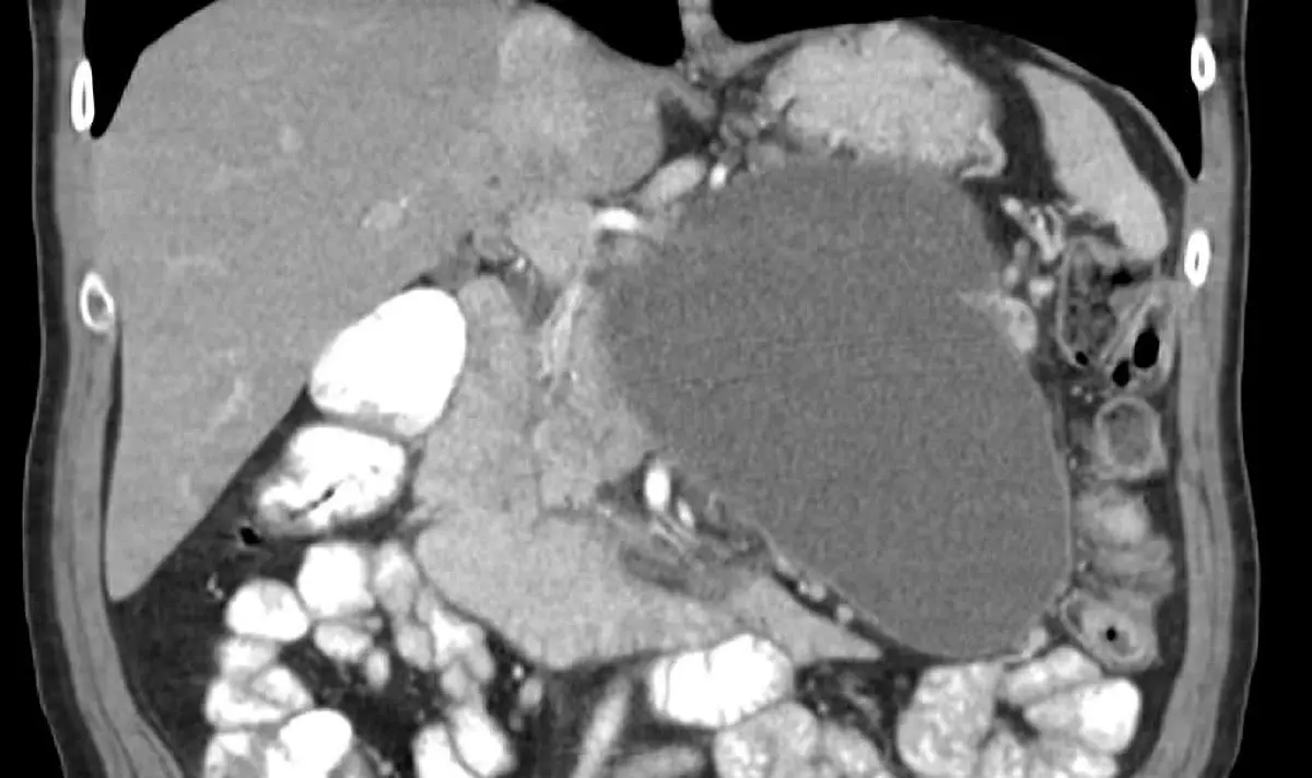
You went in for a scan because of a kidney stone, a stubborn backache, or a stomach pain that would not settle. The report came back mentioning something nobody was looking for: a cystic lesion in the pancreas, or perhaps the letters IPMN. If you have already searched for "pancreatic cyst", you have almost certainly landed on frightening content about pancreatic cancer. Here is the part that rarely reaches the top of the results: incidental pancreatic cysts are common, they grow more common with age, and the large majority are benign and never cause harm. Only a minority carry meaningful potential to turn into something serious, and those are followed or treated on well-established protocols.
What does "cystic lesion in the pancreas" mean?
A cyst is simply a fluid-filled pocket. In the pancreas, that pocket can form for very different reasons, and the reason is what decides whether it matters at all. The word "cyst" on your report describes a shape and a fluid. It is not, by itself, a diagnosis.
Most are found by accident, on a CT or MRI ordered for something else entirely. They turn up far more often than they used to, because scanners have improved and more people are scanned. Age is the clearest pattern: the older the group studied, the more cysts are found.
The main types of pancreatic cyst
The type matters far more than the word "cyst" does. Some have essentially no potential to become cancer, others have a real one, and telling them apart is the whole job.
- Pseudocyst: not a tumor at all, but a walled-off collection of fluid left behind by pancreatitis or an injury. It has no tumor lining and no potential to become cancer, and your history usually gives it away
- Serous cystadenoma: a benign tumor made of many tiny compartments, which radiologists describe as a honeycomb or microcystic appearance. It is essentially always benign, and most are simply left alone
- Mucinous cystic neoplasm (MCN): a mucin-filled cyst, usually a single cavity with a defined wall, found most often in the body or tail of the pancreas and overwhelmingly in women. It does not communicate with the duct, and because it carries genuine malignant potential over time, removal is often recommended in people fit for surgery
- Intraductal papillary mucinous neoplasm (IPMN): the type most likely to be named in your report. IPMNs grow from the lining of the pancreatic duct, produce mucin, and communicate with the duct system. They are classified by the duct involved: branch-duct (the most common, and generally the lowest risk), main-duct, and mixed type. Main-duct and mixed IPMNs carry a considerably higher risk of harboring or developing cancer, which is why the distinction is not a technicality
- Simple or true cysts: uncommon in the pancreas, thin-walled, with no solid parts. They behave much like the harmless cysts found elsewhere in the abdomen, such as a simple renal cyst on ultrasound
The features doctors actually look for
Guidelines sort the findings into so-called worrisome features, which prompt a closer look, and high-risk stigmata, which move the conversation toward surgery. In plain language, what draws attention is:
- Size: cysts of roughly 3 cm and above get more attention, though size alone is rarely the whole story
- A dilated main pancreatic duct
- An enhancing mural nodule: a solid bump on the inner wall of the cyst that takes up contrast
- A thickened, enhancing cyst wall
- Growth over time, judged against an older scan
- Jaundice (yellowing of the skin or eyes), particularly when the cyst sits in the head of the pancreas
- Unexplained weight loss, new-onset diabetes, or pancreatitis with no other cause
- A raised CA 19-9 blood test
Please do not read that as a self-diagnosis checklist. Every item can appear for reasons that have nothing to do with a cyst, and CA 19-9 in particular is a poor test in isolation. They earn their value only when read together, in context, by someone who does this work regularly: the same discipline that lets a radiologist call a hepatic hemangioma benign and leave it alone.
Symptoms, and why most cysts have none
The typical incidental pancreatic cyst produces no symptoms whatsoever, which is exactly why it arrives as a surprise, on a scan ordered to look at kidney stones on CT or an ache nobody could explain. A larger cyst can occasionally cause dull discomfort in the upper abdomen or back, nausea, or a sense of fullness after small meals. Pseudocysts usually follow a memorable bout of pancreatitis. Jaundice, unexplained weight loss, and new-onset diabetes are the symptoms clinicians take more seriously: not because they mean cancer (they very often do not) but because they deserve a proper explanation.
How pancreatic cysts are diagnosed and followed up
MRI with MRCP, a sequence that maps the pancreatic and bile ducts, is the workhorse of surveillance. It involves no radiation, and it is the best way to show whether a cyst communicates with the duct, the single most useful clue for separating an IPMN from the other types. CT is excellent in an emergency and for planning an operation, but years of repeated CT for routine monitoring is less appealing.
When a cyst has concerning features, or the imaging is genuinely ambiguous, the next step is often endoscopic ultrasound (EUS). A probe passed down into the stomach and duodenum sits millimeters from the pancreas, and fine-needle aspiration through it can sample the cyst fluid for analysis.
Two guideline sources shape most of what follows: the international (Fukuoka) consensus guidelines for IPMN and MCN, and the American College of Radiology's white paper on incidentally discovered pancreatic cysts. Both build their recommendations around cyst type, size, and features, and both accept a long horizon of monitoring for cysts that stay quiet. Intervals differ between them and are revised as evidence accumulates, so the schedule that applies to you is a conversation with your own doctor.
Treatment, and why watch and wait is a real plan
For most people the treatment is surveillance, and that is not a doctor being dismissive. Monitoring a cyst with no worrying features is an evidence-based decision, and it exists because the alternative is not free. Pancreatic surgery is major surgery: a Whipple procedure (removing the head of the pancreas along with parts of the duodenum, the bile duct, and sometimes the stomach) is among the most demanding abdominal operations there is, and even a smaller distal pancreatectomy carries a real risk of complications and of diabetes afterwards. Going through that to remove a lesion that was never going to harm you is a poor trade.
Surgery earns its place when the features genuinely point that way: a main-duct or mixed-type IPMN, an enhancing mural nodule, an MCN in a patient fit for an operation, or a cyst causing obstruction. Then the calculation flips and the operation becomes the safer path. Pseudocysts follow a different logic: most settle on their own, and drainage is reserved for those that persist, become infected, or press on nearby structures.
Why a second read can help
Characterizing a pancreatic cyst is one of the most reader-dependent tasks in abdominal imaging, and the stakes are unusually concrete. Is that bump on the cyst wall a true enhancing mural nodule, or a blob of mucin resting harmlessly against the lining? Is the main duct genuinely dilated, or at the upper end of normal? Does the cyst really communicate with the duct, or just sit beside it? Those three calls are the difference between a scan every year or two and a referral to a pancreatic surgeon, and they depend on the right MRI sequences being acquired, on prior scans being pulled up for comparison, and on someone with abdominal subspecialty experience looking closely. An independent second read of your MRI or CT can confirm the type, resolve the ambiguous features, and give you something concrete to bring to your gastroenterologist. Services like DocOrbit make that kind of expert review straightforward, and confirming that a cyst is harmless is every bit as valuable as catching one that is not. Our guide on when to get a second radiological opinion walks through the decision.
Is a pancreatic cyst always cancer?
No. The large majority of pancreatic cysts found by chance on a scan are benign and never become cancer. Some types, such as pseudocysts and serous cystadenomas, have essentially no malignant potential at all. A minority, mainly mucinous cystic neoplasms and IPMNs, carry a real but variable risk over time, which is exactly why they are characterized carefully and then followed.
What is an IPMN, and is it dangerous?
An IPMN (intraductal papillary mucinous neoplasm) is a mucin-producing growth that arises from the lining of the pancreatic duct and communicates with it. Branch-duct IPMNs are the most common and generally the lowest risk of the subtypes, while main-duct and mixed-type IPMNs carry a considerably higher risk and are more often treated surgically. Most branch-duct IPMNs without worrying features are simply monitored rather than removed.
How big does a pancreatic cyst have to be before it is a concern?
Size matters, but it is never the only thing that matters. Cysts of roughly 3 cm and above generally attract closer attention, and growth compared with an older scan is watched carefully. That said, a small cyst with an enhancing nodule or a dilated main pancreatic duct is taken more seriously than a larger, featureless one, so size is always read alongside everything else on the scan.
How often does a pancreatic cyst need to be rechecked?
Surveillance intervals depend on the type of cyst, its size, and whether it has any concerning features, and they differ somewhat between guidelines. MRI with MRCP is the usual method, because it avoids radiation and shows the duct anatomy well. For many people this becomes a long-term arrangement, and stability across several scans is genuinely good news rather than a stalemate.
Do pancreatic cysts cause symptoms?
Most do not. The typical incidental cyst is found on a scan ordered for something unrelated and causes nothing at all. Larger cysts can occasionally produce vague upper abdominal or back discomfort, nausea, or a sense of fullness. Jaundice, unexplained weight loss, new-onset diabetes, or an episode of pancreatitis are the symptoms doctors want explained rather than ignored.
Can a pancreatic cyst go away on its own?
Pseudocysts, which form after an episode of pancreatitis, often shrink or disappear without any treatment. True cystic tumors such as IPMNs, mucinous cystic neoplasms, and serous cystadenomas do not resolve on their own, though many stay the same size for years. That stability is the usual outcome, and it is one reason monitoring rather than surgery is the standard approach for low-risk cysts.
Key takeaways
- A cyst found by chance in the pancreas is a common finding, and the large majority are benign
- The type matters far more than the word cyst: pseudocysts and serous cystadenomas are essentially harmless, while MCNs and main-duct or mixed IPMNs carry real risk
- Attention goes to size, a dilated main duct, an enhancing mural nodule, a thickened wall, jaundice, weight loss, new-onset diabetes, and a raised CA 19-9
- MRI with MRCP is the workhorse for surveillance, and long-term monitoring is an evidence-based plan rather than a brush-off
If the rest of your report reads like another language, our free radiology report explainer rewrites it in everyday words, defines the terms it uses and suggests what to ask at your next appointment.
Can I ask a radiologist about my MRI report online?
Yes. Online radiology second opinions run entirely from home: you upload your images, with no CD to post and no second trip, and a subspecialist radiologist re-reads the study and writes a structured report you can give to your own doctor. DocOrbit returns that signed second read in 24 to 48 hours. You can then message the radiologist with questions about the MRI and about what the report actually means for you.
This article is for general information only and is not medical advice. Always discuss your imaging results and any next steps with a qualified physician.
