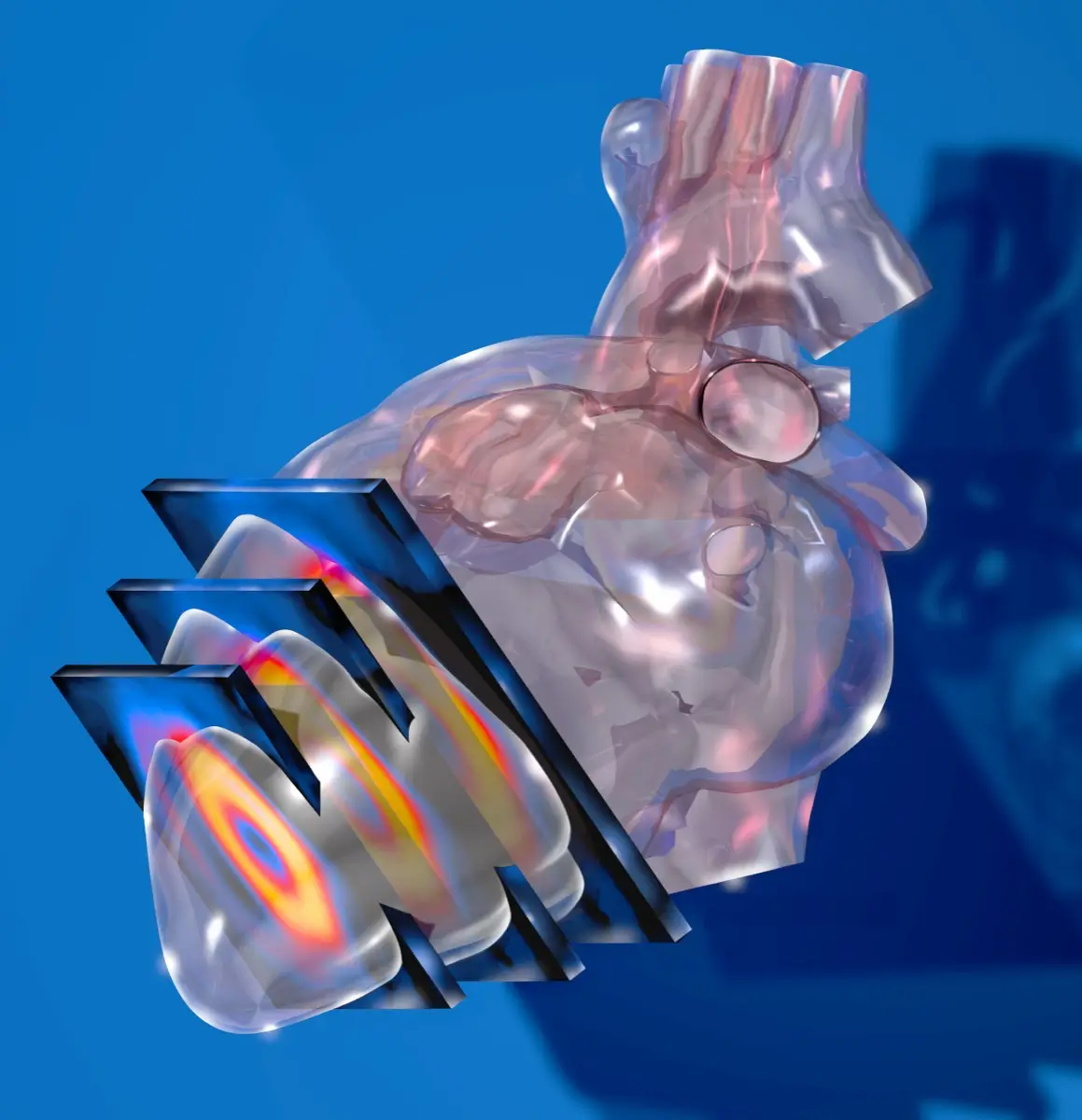
You walked on a treadmill or had a drug injected, sat under a rotating camera for twenty minutes, and now a report has come back talking about "a reversible defect in the inferior wall" or "a fixed perfusion abnormality". Here is the part nobody tells you up front: this test is deliberately designed to be sensitive, which means it flags things that turn out to be nothing far more often than it finds something dangerous. Most people who have this scan do not end up in an operating room. Below is what the words actually mean, in the order your cardiologist reads them.
What a myocardial perfusion scan actually measures
The scan does not photograph your arteries. It photographs your heart muscle and asks a simpler question: is every part of it getting enough blood?
A small amount of radioactive tracer is injected into a vein. Heart muscle with a healthy blood supply soaks it up; muscle that is being short-changed soaks up less. A gamma camera then builds a map of the whole left ventricle, and the areas that took up less tracer show up as cooler patches on the colour scale.
The trick is that the scan is done twice: once while your heart is working hard (stress) and once while it is relaxed (rest). Comparing the two pictures is where the diagnosis lives.
- Stress phase — either a treadmill or, if you cannot exercise, a drug such as regadenoson, adenosine, or dobutamine that widens your arteries or speeds your heart the way exercise would
- Rest phase — the same imaging with your heart under no particular demand
- Gated images — the camera also times the acquisition to your heartbeat, which lets it measure your pumping strength at the same time
Reversible, fixed, and what the difference really means
Almost every question patients have about this report comes down to one distinction.
A reversible defect is cool on the stress images and normal at rest. The muscle is alive and well supplied when it is idle, but the artery feeding it cannot deliver extra blood when demand rises. That pattern is the classic signature of a narrowed coronary artery, and it is what doctors call ischaemia. It is also, importantly, the pattern most likely to improve with treatment, because the muscle itself has not been damaged.
A fixed defect looks the same on both sets of images. Traditionally this suggests scar tissue from an old heart attack, sometimes one the person never knew they had. But fixed defects are also the pattern most often produced by artefact rather than disease, which is why a good reader treats them with more scepticism than patients expect.
Partial reversibility sits in between: a region that improves at rest but does not fully normalise, often meaning scar with living muscle around its edge.
The report will usually also grade the size and depth of the defect, because a small mild patch and a large deep one lead to completely different conversations.
The other numbers on the report
Two figures tend to catch the eye.
Ejection fraction (EF) is the percentage of blood the left ventricle pushes out with each beat. Roughly 50 to 70 percent is the usual normal band. A number a few points outside it is common and often means little, because this measurement genuinely varies between machines and readers.
Summed stress score, summed rest score, and summed difference score are the arithmetic behind the words. The summed difference score, in particular, is a numerical stand-in for how much reversible ischaemia is present. You do not need to interpret these yourself, but seeing them helps explain why two reports that both say "abnormal" can mean very different things.
Why some scans look abnormal when the heart is fine
This is the part that reassures people most, and it is worth understanding.
- Soft-tissue attenuation — breast tissue can dim the front wall, and the diaphragm can dim the bottom wall, producing convincing false defects
- Patient movement — even small shifts during the acquisition smear the images
- Bowel or liver uptake — bright activity next to the heart can distort the scaling of the nearby wall
- Left bundle branch block — a conduction pattern that reliably creates an apparent septal defect with no artery narrowing behind it
- Balanced three-vessel disease — the opposite problem, where everything is underperfused equally so nothing stands out as relatively cool
Good labs correct for much of this with attenuation-correction software, prone imaging, or a repeat acquisition. When a report reads "probable attenuation artefact", that is not a hedge, it is a specific and very common finding.
What usually happens next
A normal or low-risk scan is genuinely reassuring: the annual risk of a cardiac event after one is low, and most people are simply managed with risk-factor control and follow-up.
A small or mild reversible defect usually leads to medical therapy first, which means statins, blood-pressure control, sometimes an antiplatelet drug and an anti-anginal. Large-scale trials have made cardiologists more comfortable managing moderate disease this way rather than proceeding straight to a catheter.
A large or deep reversible defect, a defect in several territories, or a defect combined with a dropping ejection fraction is the combination that prompts an invasive angiogram, where a catheter shows the arteries directly and any narrowing can be treated in the same sitting.
If your scan was ordered because of calcium seen on another test, it is worth reading how those two results fit together in our piece on the coronary artery calcium score, and how the underlying process develops in atherosclerotic plaque.
What you can change
Perfusion imaging measures the consequence of coronary disease, and the inputs to that disease are unusually responsive to ordinary decisions. Stopping smoking produces measurable risk reduction within the first year. Blood pressure, LDL cholesterol, and blood sugar control each act directly on the artery wall. Regular moderate exercise, once your cardiologist has cleared you for it, improves both symptoms and outcomes. None of this is glamorous advice, but in cardiology it is the advice with the strongest evidence behind it.
Why a second read can help
Nuclear cardiology depends heavily on reader experience. Distinguishing a true inferior-wall defect from diaphragmatic attenuation, or recognising the septal pattern of a bundle branch block, is a judgement built over thousands of studies, and reader agreement in this field is good but not perfect. If your result is borderline, if it conflicts with how you actually feel, or if it is being used to justify an invasive procedure, an independent read from a subspecialist is a reasonable thing to want. DocOrbit can arrange that second read from a nuclear medicine specialist and send you a written report you can hand to your own cardiologist. Our guide on when to get a second radiological opinion covers how to judge whether your case is one of them.
Does a defect on my perfusion scan mean I need a stent
Not automatically. A perfusion defect tells your cardiologist that a region of muscle is getting less blood under stress, but the decision to place a stent depends on how large the defect is, how severe your symptoms are, and what an angiogram shows. Many people with small or mild defects are treated very effectively with medication alone. Large defects involving a lot of muscle are the ones most likely to lead to a catheter procedure.
Is a myocardial perfusion scan dangerous
It is a routine test performed millions of times a year. The tracer dose is small and clears from your body within a day or two, and the radiation is comparable to other common cardiac imaging tests. If a medication is used instead of a treadmill, it can cause a few minutes of flushing, breathlessness, or a headache, which pass quickly and can be reversed with another drug if needed.
What is a normal ejection fraction on a stress test
A left ventricular ejection fraction between roughly 50 and 70 percent is generally considered normal. Numbers slightly outside that range are common and often not meaningful on their own, because the measurement varies between techniques and between readers. Your cardiologist interprets it alongside the perfusion images, your echocardiogram, and your symptoms rather than as a standalone grade.
Can a myocardial perfusion scan be wrong
It can be misleading in both directions. Breast tissue, the diaphragm, and patient movement can create shadows that mimic a defect, and balanced disease affecting all three coronary arteries can occasionally look deceptively even. This is why borderline results are often rechecked with attenuation correction, a repeat acquisition, or a different test before any major decision is made.
What should I do while waiting for my results
Keep taking your prescribed medications and stay in touch with the team that ordered the scan. Waiting is a reasonable time to write down what your symptoms actually feel like, when they happen, and how far you can walk before they start, because that history strongly shapes how the images are interpreted. Seek urgent care for chest pain at rest, pain that lasts more than a few minutes, or breathlessness that is new and severe.
Key takeaways
- The scan compares blood flow to your heart muscle under stress and at rest; the comparison, not either image alone, carries the meaning
- Reversible means a narrowed artery limiting flow under demand, and it is the pattern most likely to respond to treatment
- Fixed usually means old scar, but is also the pattern most often caused by attenuation artefact rather than disease
- Size and depth of the defect matter far more than the mere presence of one
- Most abnormal scans lead to medication and follow-up, not to an operating room
If the rest of your report reads like another language, our free radiology report explainer rewrites it in everyday words, defines the terms it uses and suggests what to ask at your next appointment.
How do I have my nuclear medicine scan reviewed by another radiologist online?
Yes. Online radiology second opinions run entirely from home: you upload your images, with no CD to post and no second trip, and a subspecialist radiologist re-reads the study and writes a structured report you can give to your own doctor. DocOrbit returns that signed second read in 24 to 48 hours. You can then message the radiologist with questions about the nuclear medicine scan and about what the report actually means for you.
This article is for general information only and is not medical advice. Always discuss your imaging results and any next steps with a qualified physician.
