Seeing the phrase "ground-glass opacity" on a chest CT report can be unsettling, especially since the term became widely known during the COVID-19 pandemic. Here is the reassuring starting point: ground-glass opacity is a description of how a patch of lung looks on a scan, not a diagnosis, and most of the time it is caused by something temporary like an infection or mild inflammation. This article explains what radiologists mean by the term, what commonly causes it, when it matters, and how it is usually followed up.
What does "ground-glass opacity" mean?
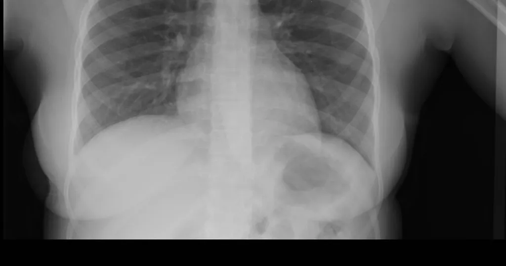
Ground-glass opacity, often shortened to GGO, describes an area of the lung that looks slightly hazier than normal on a CT scan, a little like looking through frosted or "ground" glass. The key feature is that the haze is faint enough that you can still see the normal lung markings — the blood vessels and airway walls — running through it. That is what separates it from a denser white-out called consolidation, where those structures are completely hidden.
It is almost always a CT finding rather than something seen on a plain chest X-ray, because CT is far more sensitive to these subtle differences in lung density. Radiologists describe it in several ways that carry meaning:
- Whether it is focal (a single patch) or diffuse (spread across large areas of both lungs)
- Whether it is a pure ground-glass patch or mixed with denser areas
- Whether it is rounded like a nodule or spread out in a hazy zone
- Whether it has changed compared with any earlier scans
These details often matter more than the term itself, because they help point toward a cause.
Common causes
Ground-glass opacity is one of the least specific findings in chest imaging. It is the lung's way of showing that the tiny air sacs (alveoli) are partly filled, or that their walls are slightly thickened, and many different things can do that. Common causes include:
- Infections, including viral pneumonia (COVID-19 among them), other chest infections, and atypical pneumonias
- Inflammation of the lung tissue, such as the various forms of pneumonitis
- A small amount of fluid, for example early or mild fluid backing up into the lungs
- Interstitial lung disease, a group of conditions that thicken the lung's supporting tissue
- Recent bleeding into the lung
- Scarring or healing changes left behind by a previous infection
- Less commonly, an early lung growth, including some slow-growing types of lung cancer that can begin as a small ground-glass spot
The same hazy appearance can therefore mean something as ordinary as a clearing chest infection or, in a small fraction of cases, something that deserves closer follow-up. The context — your symptoms, how the finding is distributed, and whether it changes over time — is what separates these possibilities. If your report also mentions a more defined spot, our explainer on a lung nodule found on a CT scan walks through how a single finding is put into context rather than read in isolation.
Is it serious?
For most people, ground-glass opacity is not the alarming finding it can sound like. A patch that appears during a chest infection and clears on a follow-up scan is usually a minor, self-limited thing. Widespread ground-glass opacity across both lungs, or a single patch that persists or grows over months, is taken more seriously and looked into further.
Doctors pay particular attention when the finding comes with significant breathlessness, fever, coughing up blood, or a steady decline in how the lungs are working. As with most imaging findings, interpretation depends on your symptoms, exam, and history rather than the scan alone. The pattern is a starting point for a conversation, not a verdict.
Symptoms
Ground-glass opacity itself does not cause symptoms — any symptoms come from whatever is producing it. Depending on the cause, that might mean:
- A cough, which may be dry or bring up mucus
- Shortness of breath, especially with exertion
- Fever or feeling generally unwell, when an infection is behind it
- No symptoms at all, when the finding is small or picked up by chance on a scan done for another reason
It is common for small areas of ground-glass opacity to be found incidentally, in a person who feels completely well, on a scan ordered for something unrelated.
How is it diagnosed and followed up?
Because ground-glass opacity is so non-specific, the next step is rarely dramatic. When an infection or inflammation is the likely cause, the most useful move is often simply to wait and repeat the CT scan after a few weeks, once any treatment has had time to work. A finding that clears confirms it was temporary and needs nothing more. The same patient, careful approach is used for other chest findings too — our guide to fluid around the lung shows how a finding is read in context rather than acted on in a rush.
A focal patch of ground-glass opacity that does not go away is followed differently. Radiologists may recommend periodic CT scans over months to years to confirm it stays stable, since a small number of persistent ground-glass nodules turn out to be very slow-growing early lung tumors that are highly treatable when watched and caught early. The exact schedule varies by patient and is guided by published frameworks radiologists use to weigh a finding's features against your personal risk.
Why a second read can help
Because ground-glass opacity can mean so many different things, its real value lies in how carefully it is interpreted alongside everything else about you — your symptoms, your history, and any earlier scans. A clear, expert read can help distinguish a routine, clearing infection from a finding that genuinely deserves follow-up, and it gives you something concrete to bring to your own doctor. DocOrbit offers an expert second read of your scan that you can share directly with your care team, so you can feel confident the finding has been understood in full context. If you are weighing whether to seek one, our guide on when to get a second radiological opinion may help, and you can read more broadly about the role of second opinions in radiology.
Does ground-glass opacity mean I have cancer?
In most cases, no. Ground-glass opacity is far more often caused by infection, inflammation, or a small amount of fluid than by cancer. A focal ground-glass spot that does not clear over time is sometimes watched as a possible early lung growth, but the large majority of these findings are benign and many disappear once the underlying cause settles.
Is ground-glass opacity the same as COVID-19?
No. COVID-19 pneumonia is one well-known cause of ground-glass opacity, which is why the term became familiar during the pandemic, but it is only one of many. Ordinary chest infections, inflammation, mild fluid buildup, and other lung conditions can all produce the same hazy appearance. The pattern alone cannot tell you the cause.
Can ground-glass opacity go away?
Yes, very often. When ground-glass opacity comes from an infection or short-lived inflammation, it usually fades over days to weeks as the lung heals. That is exactly why doctors often repeat a scan after a few weeks rather than acting on the first one, since a finding that clears on its own needs no further treatment.
How serious is ground-glass opacity on a CT scan?
It depends on what is causing it, how much of the lung is involved, and whether it persists. A small patch during a chest infection is usually minor and temporary, while widespread or steadily persistent ground-glass opacity is looked into more carefully. The finding is a description of how the lung looks, not a diagnosis on its own.
What is the follow-up for ground-glass opacity?
Follow-up varies by patient and cause. Ground-glass opacity thought to come from infection is often simply rechecked with a repeat scan after treatment, while a single ground-glass spot that lingers may be followed with periodic CT scans to make sure it stays stable. Your doctor decides the timing based on your symptoms, history, and the look of the finding.
Key takeaways
- Ground-glass opacity is a hazy area on a CT scan where you can still see the lung's normal markings. It is a description, not a diagnosis
- It is highly non-specific: infections, inflammation, mild fluid, and old scarring are far more common causes than cancer
- COVID-19 is only one of many possible causes, despite how familiar the term became during the pandemic
- Many cases clear on their own, which is why a repeat scan after a few weeks is a common next step
- A focal patch that persists may be followed with periodic scans, and interpretation always depends on your symptoms and history
This article is for general information only and is not medical advice. Always discuss your imaging results and any next steps with a qualified physician.