Reading "degenerative disc disease" on a spine MRI report can sound frightening — the word disease makes it seem like something is seriously wrong, or that your back is wearing out ahead of schedule. Here is the reassuring truth to start with: for most people it is not really a disease at all, but the normal, expected way the spinal discs change as we age. It is one of the most common findings on a back or neck MRI, and on its own it rarely means anything is broken. Here is what the term actually describes, why it shows up, and when the back pain around it is worth a closer look.
What does "degenerative disc disease" mean?
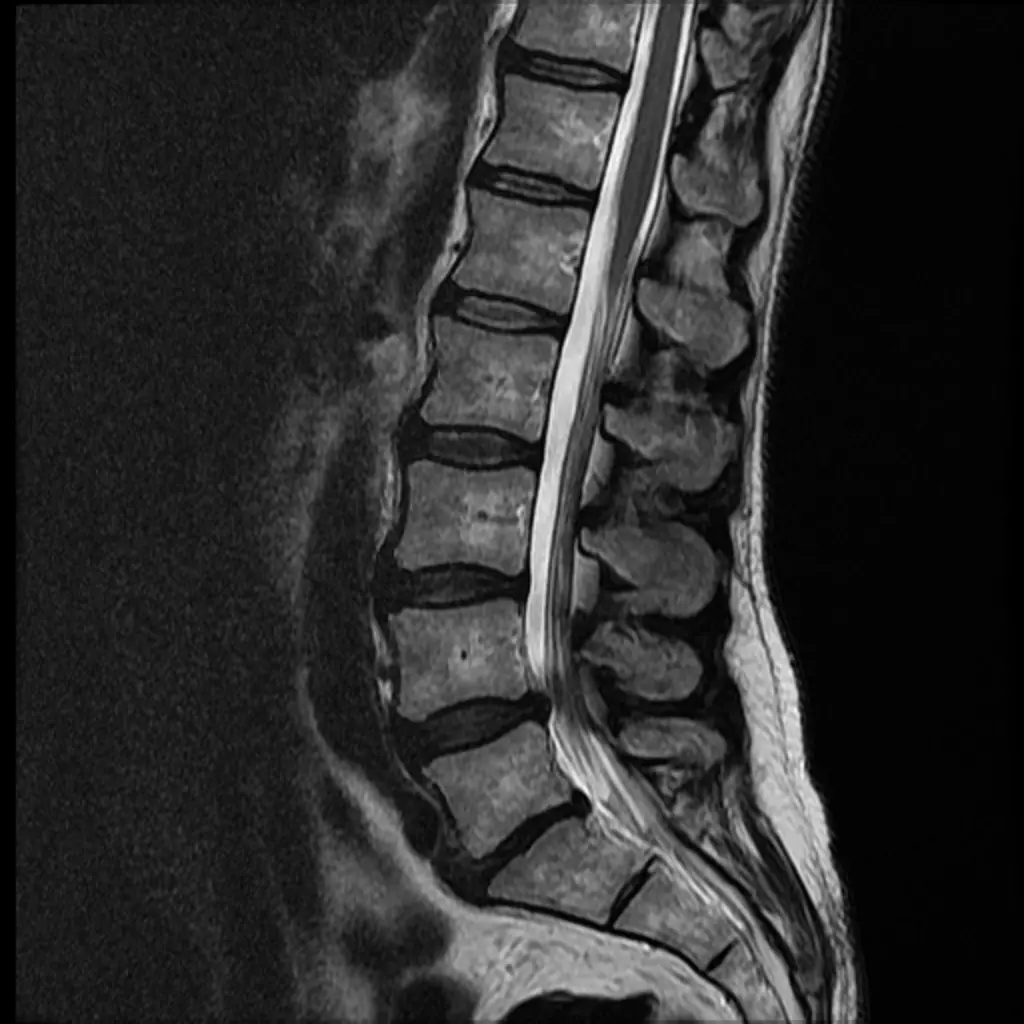
Between each pair of vertebrae in your spine sits a disc — a tough, rubbery cushion with a soft, water-rich center that works as a shock absorber. Over the years those discs naturally lose some of their water content, flatten a little, and become less springy. On an MRI, a radiologist sees this as discs that look darker than usual on certain sequences, slightly reduced in height, or bulging gently at the edges. That collection of age-related changes is what gets labeled degenerative disc disease.
Despite the name, it is not an infection or a progressive illness in the way we usually think of disease. Radiologists often describe what they see in layers:
- Disc desiccation — the disc has dried out and lost water, showing up darker on T2 MRI images
- Loss of disc height — the cushion has thinned, narrowing the space between two vertebrae
- Disc bulge or small protrusion — the disc edge extends slightly beyond its normal border
- Endplate and adjacent bone changes (sometimes called Modic changes) — reactions in the bone next to the disc
When a disc pushes out far enough to press on a nerve, the report may switch to a different term — you can read more in our guide to disc herniation on MRI.
What causes degenerative disc disease?
The single biggest driver is simply time. By middle age, the large majority of people have at least some disc degeneration visible on imaging, whether or not they feel any pain. Other factors can speed it up or make it more noticeable:
- Age — the most important factor by far
- Genetics — a strong family tendency toward earlier disc changes
- Smoking — reduces the blood supply to the discs and accelerates drying out
- Repeated heavy lifting or physically demanding work
- Carrying extra weight, which adds sustained load to the lower back
- A previous back injury
Because these changes are so common, finding them on a scan does not mean you did something wrong or that your spine is failing. Much like a rotator cuff tear found on a shoulder MRI, disc degeneration is often just the normal signature of a body that has been used for decades.
Is degenerative disc disease serious?
For most people, no. Studies that scan people with no back pain at all routinely find disc degeneration — one reason radiologists and spine doctors treat it as a background finding rather than an alarm. The image describes the state of the disc; it does not measure your pain or predict your future.
It becomes more meaningful when the imaging lines up with your symptoms — for example, degeneration at a level where a disc is also pressing on a nerve, which can cause pain, numbness, or weakness travelling down a leg or arm. A few findings do prompt a doctor to look more carefully:
- Pain radiating down a leg or arm, or numbness, tingling, or weakness in a limb
- Back pain that follows significant trauma
- Loss of bladder or bowel control, or numbness around the groin — this needs urgent medical attention
- Fever, unexplained weight loss, or a history of cancer alongside new back pain
These do not describe most people with degenerative discs, but they are the situations where the finding stops being routine and gets acted on quickly.
What symptoms does it cause?
Here is the part that surprises many patients: degenerative disc disease frequently causes no symptoms at all. Plenty of people learn they have it only because they had a scan for an unrelated reason.
When it does cause trouble, the common pattern is:
- Low back or neck ache that comes and goes, often worse with sitting, bending, or twisting
- Stiffness, especially in the morning or after long periods in one position
- Flare-ups that settle down over days to weeks
- If a nerve is involved, pain or tingling spreading into an arm or leg
The amount of degeneration on the scan often does not match the amount of pain a person feels. Someone with dramatic-looking images can be perfectly comfortable, while someone with mild changes can have real discomfort. That is why treatment is guided by how you feel and function — not by the MRI alone.
How is it diagnosed and followed up?
Degenerative disc disease is usually first spotted on an MRI, which shows the discs, nerves, and soft tissue in detail. X-rays and CT can reveal narrowed disc spaces and bone changes but tell you less about the disc itself. The scan is only one input — your doctor combines it with your history, a physical exam, and sometimes nerve tests to build the full picture.
For uncomplicated degenerative changes, there is usually no fixed follow-up imaging schedule. Repeat scans are ordered when symptoms change meaningfully — new leg weakness, pain that is not improving, or before a planned procedure — rather than simply to watch stable discs get older.
How is it treated?
For the large majority of people, treatment is conservative and aimed at controlling symptoms and keeping the spine strong and mobile — not at "fixing" the disc, which does not need fixing when it is quietly aging.
- Staying active — prolonged bed rest tends to make back pain worse, not better
- Physical therapy to strengthen the core and back muscles that support the spine
- Over-the-counter pain relievers or anti-inflammatories for flare-ups, as advised by a doctor
- Heat, gentle stretching, and simple posture adjustments
- Occasionally, targeted injections when nerve-related pain is limiting
Surgery is reserved for a small minority — usually when there is clear nerve compression with progressive weakness, or pain that has not responded to months of conservative care. Most people never reach that point.
Lifestyle changes that help
Because disc health is tied to your overall physical condition, everyday habits make a real difference:
- Regular low-impact exercise — walking, swimming, cycling — to keep the discs nourished and the muscles supportive
- Not smoking, which protects the blood supply feeding the discs
- Keeping a healthy weight to reduce the daily load on your lower back
- Lifting with your legs and avoiding sudden twisting under load
- Setting up a supportive chair and screen height if you sit for work
None of these reverse existing changes, but together they help keep symptoms low and function high.
Why a second read can help
Spine MRIs are detailed, and the same images can be described in very different words depending on who reads them — one report may frame everything as routine age-related change, while another lists every minor bulge in language that sounds alarming. Because that wording shapes how worried you feel and what happens next, a clear second read can add real peace of mind. DocOrbit provides an expert second opinion on your imaging that you can share with your own doctor, helping you understand whether your findings are the ordinary kind or the kind that genuinely warrants a closer look. If you are weighing whether it is worth it, our guide on when to get a second radiological opinion walks through the situations where it helps most.
Is degenerative disc disease serious?
For most people it is not. Disc degeneration is a normal, age-related change found on the majority of adult spine scans, and it often causes no symptoms. It becomes more important when the imaging matches symptoms such as pain, numbness, or weakness spreading into a limb, which is when a doctor looks more closely.
Does degenerative disc disease always get worse over time?
Not in a way that necessarily matters. The disc changes themselves tend to progress slowly with age, but pain does not track the images closely. Many people find their symptoms stay stable or even improve with exercise and good habits, even as the discs continue to age.
Can degenerative disc disease be reversed?
The structural changes in a disc cannot be reversed, and no supplement or treatment restores a disc to its younger state. The good news is that reversing the images is not the goal. Most people control symptoms well and stay active through exercise, weight management, and physical therapy without ever changing what the MRI shows.
What is the best treatment for degenerative disc disease?
For the large majority, the best approach is conservative: staying active, physical therapy to strengthen the muscles that support the spine, and short-term pain relief during flare-ups. Surgery is reserved for the small number of people with nerve compression and progressive weakness, or pain that has not responded to months of other treatment.
Should I avoid exercise if I have degenerative disc disease?
No, in most cases the opposite is true. Prolonged rest tends to make back pain worse, while regular low-impact exercise keeps the supporting muscles strong and the discs nourished. Your doctor or physical therapist can help you choose activities that are safe for your specific situation.
Key takeaways
- Degenerative disc disease is usually normal, age-related wear — not a disease in the way the name suggests
- It is one of the most common spine MRI findings and frequently causes no symptoms at all
- The amount of degeneration on the scan does not predict the amount of pain you feel
- Most people are managed well by staying active, exercising, and doing physical therapy — surgery is rare
- See a doctor promptly for leg or arm weakness, spreading numbness, or any loss of bladder or bowel control
This article is for general information only and is not medical advice. Always discuss your imaging results and any next steps with a qualified physician.