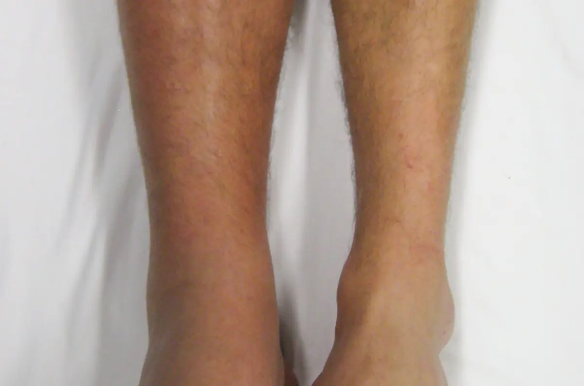
Seeing the words "deep vein thrombosis," "DVT," or "venous thrombosis" on an ultrasound report, or hearing that you have a blood clot in your leg, is understandably frightening. Here is the balanced truth: a DVT is a common and highly treatable condition, and when it is caught and treated promptly, the large majority of people recover well. It is also one finding you should not ignore, because a clot in a deep vein deserves proper medical attention. This article explains what a DVT actually is, why clots form, the symptoms to watch for, when it becomes an emergency, and how it is treated.
What is a deep vein thrombosis?
A deep vein thrombosis is a blood clot that forms in one of the deep veins, the larger veins that run deep inside the body, most often in the calf or thigh. Less commonly, a clot can form in the veins of the pelvis or, rarely, in an arm.
Your legs have two sets of veins: superficial veins just under the skin, and deep veins buried within the muscles. A clot in a superficial vein (called superficial thrombophlebitis) is usually less serious. A clot in a deep vein is what doctors mean by DVT, and it matters more because of where it sits and where it can travel.
A DVT is a problem of the veins, the vessels that carry blood back to the heart. That makes it different from arterial conditions such as atherosclerotic plaque, which builds up in the arteries that carry blood away from the heart.
On a scan, DVT is most often confirmed with a Doppler or compression ultrasound of the leg. The person performing the scan presses gently on the vein with the probe: a healthy vein squashes flat, while a vein filled with clot does not compress. They also check the blood flow through the vein, which is reduced or absent when a clot is blocking it. Typical signs include:
- A vein that does not compress under gentle pressure, the key sign of a clot
- Reduced or absent blood flow on Doppler imaging
- Sometimes, visible clot material inside the vein
What causes a blood clot in the leg?
Clots tend to form when one or more of three things happen: blood flows too slowly, the vein wall is injured, or the blood itself clots more easily than it should. Doctors call this combination Virchow's triad. In everyday terms, the common triggers and risk factors include:
- Long periods of not moving, such as a long flight or car journey, bed rest, or recovery after surgery
- A recent surgery or injury, especially to the hip, knee, or leg
- Pregnancy and the weeks after giving birth, when blood clots more readily
- Estrogen-containing birth control pills or hormone replacement therapy
- Cancer and some cancer treatments
- Being significantly overweight, smoking, or older age
- Inherited conditions that make the blood clot more easily (thrombophilias), such as Factor V Leiden
- Dehydration, and having had a clot before
Often more than one factor lines up at the same time, for example a long flight taken shortly after an operation.
What are the symptoms of a DVT?
A DVT usually affects one leg, and the classic symptoms come from blood backing up behind the clot:
- Swelling in one leg, often the calf, that does not settle
- Pain or tenderness, frequently in the calf, that can feel like a cramp or a deep ache
- Warmth over the affected area
- Skin that looks red, discolored, or slightly shiny and stretched
Not every DVT causes obvious symptoms, and some are found only when a scan is done for another reason. Equally, a swollen or sore calf has many harmless causes, such as a muscle strain, so symptoms alone cannot confirm or rule out a clot, which is exactly why imaging is used.
Is a DVT serious, and when is it an emergency?
This is the part worth being clear about. A DVT is taken seriously mainly because a piece of the clot can break off and travel through the bloodstream to the lungs, where it becomes a pulmonary embolism. A large pulmonary embolism is a genuine medical emergency. That is the reason doctors treat a confirmed DVT promptly rather than waiting to see what happens.
The reassuring side is that with prompt treatment, the risk of a clot traveling drops substantially, and most people never develop a serious complication.
Seek emergency care right away if you have any of the following, which can signal that a clot has reached the lungs:
- Sudden shortness of breath
- Chest pain that may worsen when you breathe in
- Coughing, sometimes with blood
- A racing heartbeat or feeling faint
These symptoms need immediate attention, not a wait-and-see approach.
How is a DVT diagnosed?
If a DVT is suspected, doctors usually combine a few steps. They start by weighing your risk factors and symptoms, sometimes using a simple scoring system. A blood test called a D-dimer can help: a normal result makes a clot unlikely, while a raised result means imaging is needed to be sure.
The main imaging test is a compression or Doppler ultrasound of the leg, which is quick, painless, and uses no radiation. For clots suspected higher up in the pelvis or abdomen, where ultrasound is harder, a CT venogram or MR venogram may be used instead. Because ultrasound findings depend partly on the person performing the scan, borderline results are sometimes repeated after a few days or reviewed again for a closer look.
How is a DVT treated?
The mainstay of treatment is anticoagulant medication, often called blood thinners. These do not dissolve the existing clot directly; instead they stop it from growing and prevent new clots from forming while your body gradually breaks the clot down on its own. Commonly used options include direct oral anticoagulants (DOACs) such as rivaroxaban or apixaban, and in some situations heparin or warfarin.
Most people take blood thinners for at least three months; the exact length depends on why the clot formed and your personal risk of another one. Some people also wear graduated compression stockings to reduce swelling and lower the risk of longer-term leg symptoms.
Modern advice is usually to keep moving rather than resting in bed once treatment has started, because gentle activity supports healthy blood flow. In more severe cases, doctors occasionally use clot-dissolving treatment (thrombolysis) or a procedure to remove the clot, and for people who cannot take blood thinners, a small filter can be placed in the main abdominal vein to catch clots before they reach the lungs. These are the exception rather than the rule.
Can you lower your risk of a DVT?
Several everyday steps genuinely reduce the risk, especially if you already have risk factors:
- On long flights or drives, move your legs regularly, walk when you can, and stay hydrated
- Get up and move as soon as it is safe after surgery or illness, following your care team's advice
- If you are at higher risk, ask about compression stockings or preventive blood thinners around surgery or long travel
- Keep a healthy weight, stay active, and stop smoking, all of which support healthy circulation
- Tell your doctor about any personal or family history of clots before starting hormone therapy or major surgery
Why a second read can help
A DVT is usually diagnosed on ultrasound, and ultrasound is more operator-dependent than most other scans, meaning the result can hinge on how and when the study was done. Telling an acute clot from an old, healed one, or judging a borderline result in a calf vein, is exactly the kind of call where a careful second read adds confidence. An expert second opinion, such as the one DocOrbit provides on your images and report, can confirm the finding and its extent so you and your own doctor can be sure of the plan. If you are weighing whether it is worth it, our guide on when to get a second radiological opinion walks through the situations where it matters most.
Is a DVT a medical emergency?
A DVT itself is usually treated urgently but is not always an emergency on its own. It becomes an emergency if part of the clot travels to the lungs, causing a pulmonary embolism. Seek immediate care for sudden shortness of breath, chest pain, or coughing up blood.
Can a DVT go away on its own?
The body can slowly break down a clot over time, but a deep vein clot should not be left untreated because of the risk that it travels to the lungs. Treatment with blood thinners is used to stop the clot from growing and to prevent new clots while healing takes place.
How long does it take for a leg clot to dissolve?
Blood thinners usually start lowering the risk of the clot spreading within days, but the clot itself can take weeks to months for the body to break down. Most people take anticoagulants for at least three months, and some for longer depending on the cause.
Can I walk with a DVT?
In most cases gentle walking is encouraged once treatment has begun, because staying active supports healthy blood flow. Prolonged bed rest is no longer the standard advice. Always follow the specific guidance of the doctor treating you.
What happens if a DVT is left untreated?
An untreated DVT carries a higher risk of the clot growing or breaking off and traveling to the lungs as a pulmonary embolism, which can be dangerous. Untreated clots can also lead to long-term leg swelling and discomfort known as post-thrombotic syndrome. This is why prompt treatment matters.
Key takeaways
- A DVT is a blood clot in a deep vein, most often in the calf or thigh, usually confirmed with a leg ultrasound
- It is common and highly treatable, but it should never be ignored, because a clot can travel to the lungs
- Warning signs of a clot reaching the lungs, such as sudden breathlessness or chest pain, need emergency care
- Blood thinners are the main treatment, usually for at least three months, and gentle movement is encouraged
- Staying mobile and hydrated during long travel and after surgery is one of the best ways to lower your risk
Still unsure what the rest of your report says? Paste it into our free radiology report explainer and read it back in plain English, with each medical term defined and a short list of questions worth asking your doctor.
Can I ask a radiologist about my ultrasound report online?
Yes. Online radiology second opinions run entirely from home: you upload your images, with no CD to post and no second trip, and a subspecialist radiologist re-reads the study and writes a structured report you can give to your own doctor. DocOrbit returns that signed second read in 24 to 48 hours. You can then message the radiologist with questions about the ultrasound and about what the report actually means for you.
This article is for general information only and is not medical advice. Always discuss your imaging results and any next steps with a qualified physician.
