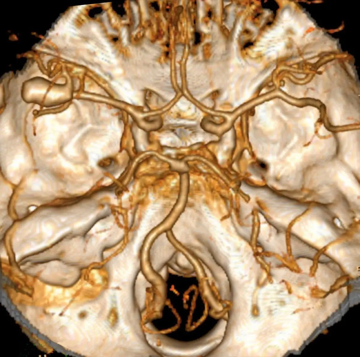
Few phrases on a brain scan report unsettle people like "aneurysm", especially when it turns up unexpectedly on an MRI or CT done for headaches, dizziness, or something entirely unrelated. Here is the reassuring place to start: the large majority of brain aneurysms are small, unruptured, and quietly stable, many are found completely by accident, and a great many never cause any trouble at all. This article explains what an aneurysm actually is, why they form, how doctors judge which ones matter, the warning signs that count, and what treatment looks like when it is needed.
What a brain aneurysm actually is
A brain aneurysm, sometimes called a cerebral or intracranial aneurysm, is a small bulge or balloon that forms at a weak spot in the wall of an artery in or around the brain. Most develop where arteries branch, because those junctions take the most mechanical stress over a lifetime. The most common type is a "saccular" or "berry" aneurysm, named because it looks like a small berry hanging from the vessel.
On imaging, an aneurysm shows up as a rounded outpouching from an artery. It is usually seen best on studies that map the blood vessels directly: CT angiography (CTA), MR angiography (MRA), or catheter angiography (DSA), which remains the most detailed. A standard MRI or CT of the head can sometimes hint at one, which is often how an incidental aneurysm is first spotted before a dedicated vessel study confirms it.
- "Unruptured" means the aneurysm has not bled, which is the situation for most that are found
- "Saccular" or "berry" describes the common rounded shape
- "Fusiform" describes a less common, more spindle-shaped widening of the vessel
- The report usually gives a size in millimeters and a location, both of which guide the plan
How common are they, and what causes them
Unruptured brain aneurysms are more common than most people expect. Estimates suggest that roughly 1 in 50 adults carries one, and the great majority live their whole lives without ever knowing, because the aneurysm never ruptures and never causes symptoms. Finding one on a scan is far more often the start of a monitoring plan than the start of bad news.
Aneurysms form from a mix of factors that weaken an artery wall over time. Some are within your control, which is part of why the finding can be useful information.
- High blood pressure — a major, and very treatable, contributor to arterial wall stress
- Smoking — one of the strongest modifiable risk factors for both formation and rupture
- Family history — having close relatives with aneurysms raises the odds
- Age and sex — they are found more often with increasing age and somewhat more often in women
- Certain inherited conditions — such as polycystic kidney disease and some connective-tissue disorders
- Heavy alcohol use and stimulant drugs — which raise blood pressure
The same vascular wear that drives aneurysm formation overlaps with the arterial changes we describe in our explainer on atherosclerotic plaque on a scan, and controlling blood pressure helps both.
Is it serious? How doctors judge the risk
This is the honest heart of the matter: an unruptured aneurysm is not an emergency, but it is a finding doctors take seriously and assess carefully. The concern is rupture, which can cause bleeding around the brain called a subarachnoid hemorrhage. That is a serious event, but it is also uncommon for small, stable aneurysms, and the point of the assessment is to identify the small number that genuinely warrant treatment.
Specialists weigh several features together rather than reacting to any single one:
- Size — larger aneurysms carry a higher rupture risk; very small ones in the front circulation carry a low yearly risk
- Location — aneurysms in the back (posterior) circulation tend to be treated more readily
- Shape — irregular, lobulated, or "daughter-sac" aneurysms are watched more closely than smooth, round ones
- Growth over time — an aneurysm that enlarges on follow-up imaging is taken more seriously
- Your factors — age, smoking, blood pressure, family history, and whether you have ever had a bleed before
Doctors sometimes use structured tools, such as the PHASES score, to help estimate rupture risk and frame the conversation. These are guides for a shared decision, not automatic verdicts, and your own history always shapes the plan.
Symptoms, and the emergency you should never ignore
Most unruptured aneurysms cause no symptoms whatsoever, which is exactly why so many are found by accident. A larger aneurysm can occasionally press on nearby structures and cause a persistent headache, a drooping eyelid, double vision, or a dilated pupil, and those warrant prompt, though not necessarily emergency, evaluation.
A rupture is entirely different, and it is the one scenario to know cold. The hallmark is a sudden, explosive headache that many describe as the worst of their life, reaching full intensity within seconds. It may come with a stiff neck, nausea and vomiting, light sensitivity, brief loss of consciousness, or a seizure. This is a life-threatening emergency: anyone with these symptoms should call emergency services immediately. A so-called sentinel or warning headache can sometimes precede a major bleed, so a sudden severe headache unlike any before should never be brushed off.
How it is monitored and treated
For many small, low-risk aneurysms, the recommended plan is watchful monitoring: periodic imaging with MRA or CTA to make sure the aneurysm is not growing or changing, combined with aggressive control of blood pressure and stopping smoking. Doing nothing invasive is a legitimate, evidence-based choice when the risk of treatment would outweigh the low risk of rupture.
When treatment is advised, there are two main routes, and a specialized neurovascular team chooses between them based on the aneurysm's anatomy:
- Endovascular coiling or flow diversion — performed from inside the blood vessel through a thin catheter, with no open skull incision; tiny coils or a stent-like device redirect blood flow so the aneurysm seals off
- Microsurgical clipping — an operation in which a small metal clip is placed across the neck of the aneurysm to close it off from the circulation
Both are well-established, and each has situations where it is the better fit. Lifestyle steps matter regardless of the path chosen: keeping blood pressure well controlled and stopping smoking are among the most powerful things you can do, echoing the vascular-health themes in our piece on brain-imaging findings and what they mean.
Why a second read can help
An aneurysm sits in a genuine grey zone, where the same scan can reasonably lead to either careful monitoring or treatment depending on how millimeter-level measurements, shape, and your personal risk are weighed. That is exactly the kind of decision where a second expert perspective adds real value. DocOrbit offers an expert second read of your brain imaging that you can share with your own neurologist or neurosurgeon, which can bring clarity and confidence before you commit to watching or treating. It is another specialist set of eyes on the same pictures, never a replacement for your treating team.
Is a brain aneurysm always dangerous?
No. Most brain aneurysms are small, unruptured, and never cause a problem, and many are discovered by chance on a scan done for another reason. The risk that matters is rupture, which is uncommon for small, stable aneurysms. Whether an aneurysm needs treatment or simply monitoring depends on its size, shape, location, and your own risk factors, which is why the finding is assessed by a specialist rather than judged by the word alone.
What size of brain aneurysm is dangerous?
Size is one of the strongest predictors of rupture, and in general the larger the aneurysm, the higher the risk. Very small aneurysms under about 7 mm, especially in the front part of the brain's circulation, have a low yearly rupture risk and are often monitored. Larger aneurysms, irregular ones, or those in certain locations are more likely to be treated. Size is only part of the picture, though, so decisions are always individualized.
Can a brain aneurysm be treated without open surgery?
Often, yes. Many aneurysms today are treated from inside the blood vessel using a thin catheter threaded up from an artery, a technique called endovascular coiling or flow diversion, with no open incision in the skull. The alternative is microsurgical clipping, in which a tiny clip is placed across the neck of the aneurysm. Which approach is best depends on the aneurysm's size, shape, and location, and is decided by a specialized neurovascular team.
What are the warning signs of a ruptured brain aneurysm?
The classic warning sign is a sudden, extremely severe headache, often described as the worst headache of your life, that peaks within seconds. Other signs include a stiff neck, nausea and vomiting, sensitivity to light, double vision, a drooping eyelid, seizures, or loss of consciousness. A ruptured aneurysm is a medical emergency, so anyone with these symptoms should call emergency services immediately rather than wait.
Key takeaways
- Most brain aneurysms are small, unruptured, and found by accident, and never cause symptoms
- The risk that matters is rupture, which is uncommon for small, stable aneurysms
- Size, shape, location, growth, and your own risk factors decide whether to monitor or treat
- A sudden, worst-ever headache can signal a rupture and is a call-emergency-services emergency
- Treatment can often be done from inside the vessel by coiling, or by surgical clipping
Your report probably contains more than this one finding. Our free radiology report explainer puts the whole thing into plain English and gives you a short list of questions to take to your doctor.
Can I ask a radiologist about my scan report online?
Yes. Online radiology second opinions run entirely from home: you upload your images, with no CD to post and no second trip, and a subspecialist radiologist re-reads the study and writes a structured report you can give to your own doctor. DocOrbit returns that signed second read in 24 to 48 hours. You can then message the radiologist with questions about the scan and about what the report actually means for you.
This article is for general information only and is not medical advice. Always discuss your imaging results and any next steps with a qualified physician.
